
Sandy Weltan
The description of a histiocytoma in a well known pathology textbook is as follows:
“Cutaneous histiocytoma is a benign skin tumour that originates from the epidermal Langerhans cell and is predominantly a solitary tumour of young dogs that undergoes spontaneous regression. Histiocytoma cells may migrate to regional lymph nodes; lymphadenopathy will regress along with the primary lesion.”
In practice, lymphadenopathy is rarely seen, so this one needs special mention.
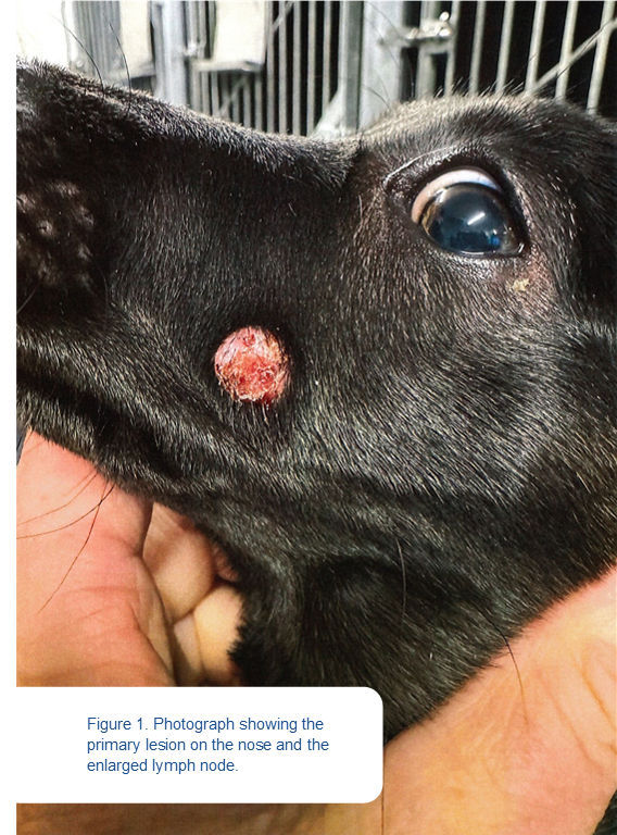
A 12-week old, female terrier presented with two lesions. One was a 1cm raised round ulcerated firm lump on left bridge of nose, which had been there approximately two weeks. The other was a 2cm diameter spherical soft lump at left angle of jaw which had been noticed two days prior (Figure 1).
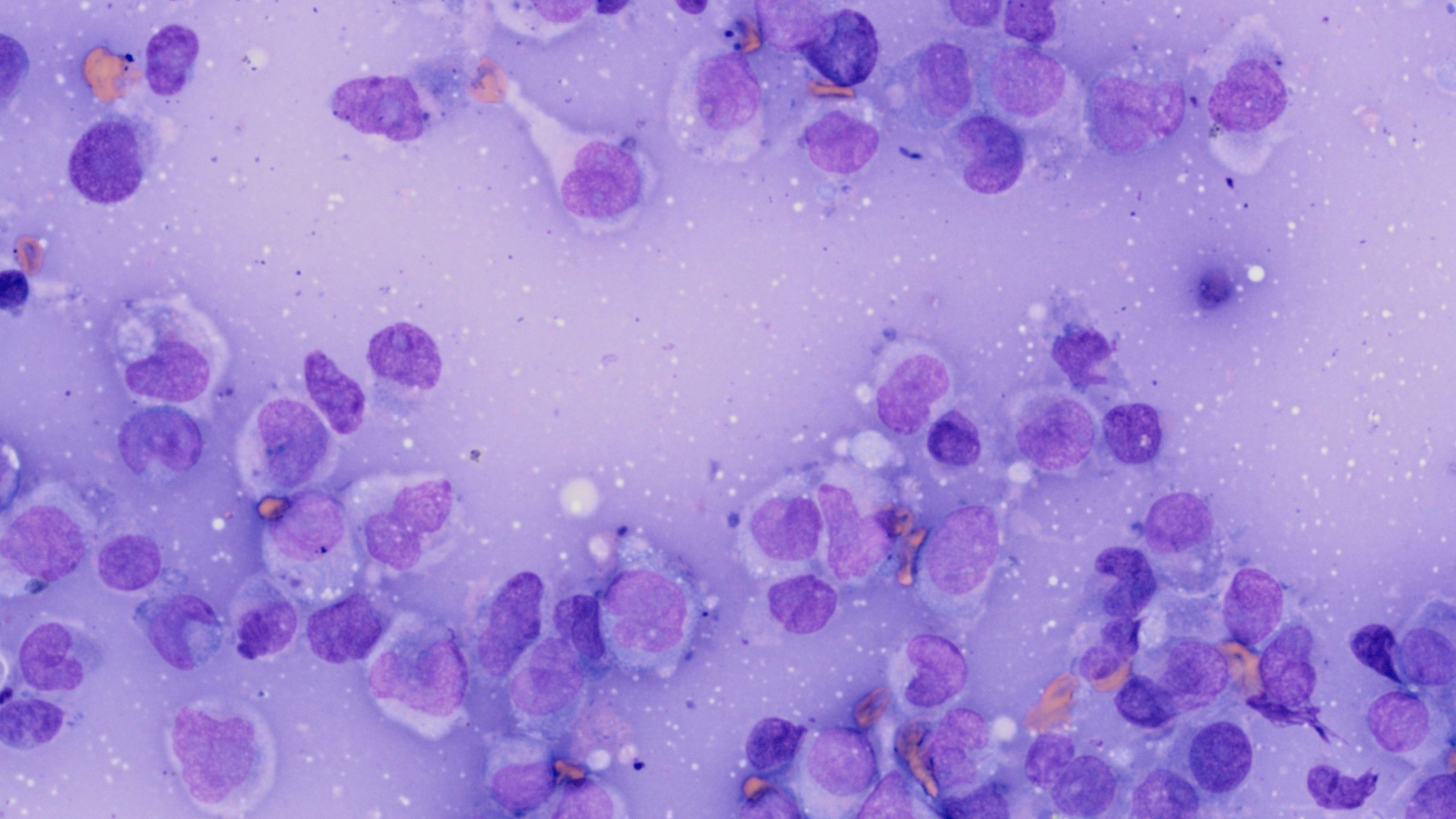
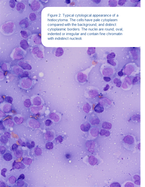
Fine needle aspirates were submitted from both masses. In the smears from the nose, the background consisted of homogenous, moderately basophilic material and the cellularity was high in most of the smears with variable cell preservation. The smear with the best cell preservation consisted of round cells which had a moderate amount of pale basophilic cytoplasm with distinct cytoplasmic borders. The nuclei were round, oval, irregular or indented and contained finely stippled chromatin with indistinct nucleoli. Small numbers of cytoplasmic fragments were present in the background with occasional small lymphocytes (Figure 2).
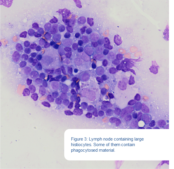
In the smears from mass at the angle of the jaw, there was a mixed population of lymphocytes and plasma cells with small, medium and large lymphocytes. There were also large numbers of histiocytic cells. They showed marked moderate pleomorphism and had a moderate amount of pale to mildly basophilic and variably vacuolated cytoplasm with distinct cytoplasmic borders.
The nuclei were mostly oval to indented and some of the cells were cytophagic, containing phagocytosed lymphocytes (Figure 3).
The highest incidence of cutaneous histiocytoma is in dogs under two years of age. Histiocytomas most commonly occur on the head, with the pinna being the most frequently affected site, although tumours can occur on many other areas of the body. Infiltration by small lymphocytes, which are cytotoxic CD8+ T cells, signals the beginning of spontaneous regression. The incidence of spontaneous regression decreases with increasing age and in dogs over three years of age, other round cell tumours need to be considered as differential diagnoses. In truth, the incidence of migration of histiocytoma cells to lymph nodes is uncommon, so this was an unusual case.
References:
1. Goldschmidt MH, Goldschmidt KH: Tumours of the skin and soft tissues. In: Tumours in domestic animals. 4 edn. Edited by Meuten DJ. Ames, Iowa: Blackwell; 2017: 315-377.
2. Raskin RE: Skin and Subcutaneous Tissues. In: Canine and Feline Cytology A Colour Atlas and Interpretation Guide. 3 edn. Edited by Raskin RE, Meyer DJ. St Louis: Elsevier; 2016: 34-90.
3. Moore PF, Schrenzel MD, Affolter VK, Olivry T, Naydan D: Canine cutaneous histiocytoma is an epidermotropic Langerhans cell histiocytosis that expresses CD1 and specific beta 2-integrin molecules. Am J Pathol 1996, 148(5):1699-1708.