

KAREN BAILEY
Clinical history:
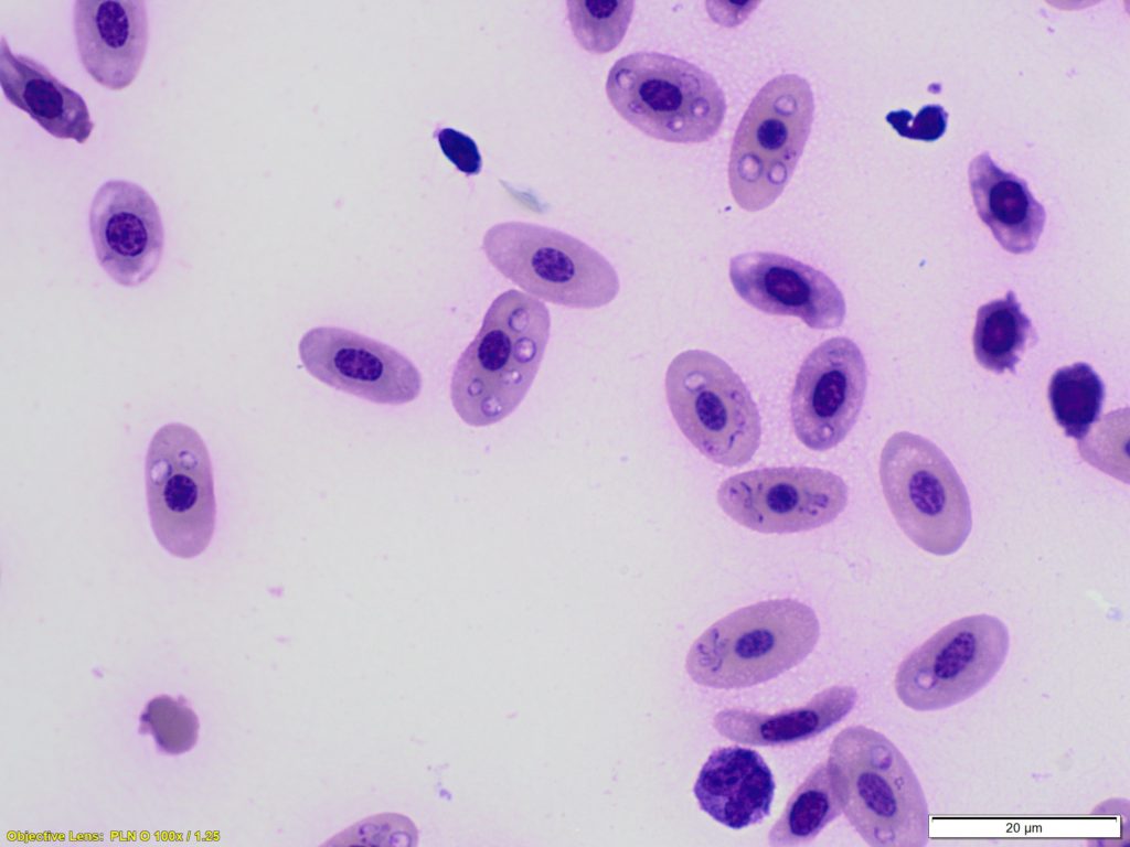
An adult male kororā / little blue penguin (Eudyptula minor) was brought to a wildlife hospital. On examination it was severely underweight at 805g (should be 1 – 1.2 kg) and had white mucous membranes. The PCV was 6% (normal 38-52%). An image from a blood smear (100x objective, 1000x magnification) is shown below.
Laboratory findings:
Very large numbers of round (signet ring) or teardrop shaped intracytoplasmic structures can be seen within the erythrocytes. These are consistent with Plasmodium species trophozoites, supporting a diagnosis of avian malaria.
Discussion:
Avian malaria is a mosquito borne protozoal disease which can affect many bird species, but is especially problematic in penguins, particularly in captive populations but also in the wild. In this case the bird died.
Cytological examination of a smear from the spleen showed 50 -75% of erythrocytes contained 1-8 haemoparasites most consistent with Plasmodium sp. (though gametocytes of Haemoproteus sp. or piroplasms from Babesia sp. could not be ruled out based on morphology alone). Histopathological examination showed haemosiderosis in the spleen, cholestasis in the liver and intratubular haemoglobin of the kidney, consistent with a haemolytic process and the most common reason for this in penguins is avian malaria.
Clinical signs in infected birds may range from nil to sudden death and include pallor, lethargy, dyspnoea, anorexia, vomiting and more rarely, neurological signs. Physiological and environmental factors such as moult, chick rearing, weather and in captive environments, husbandry, may be associated with severity of disease.
Thanks to Pauline Howard for this interesting case.