
LISA HULME-MOIR
This recent case highlights the importance of blood film examinations and illustrates how diagnoses may be missed if relying on in-clinic analyser counts alone.
Clinical history:
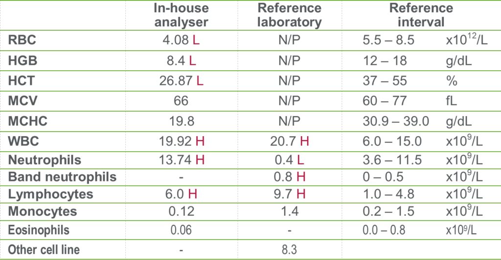
A blood sample from a 9-year-old male Labrador cross was submitted for blood film examination after presenting lethargic, off his food and with a temperature of 40.5°C. His popliteal lymph nodes were slightly enlarged but no other abnormalities were noted on physical examination. In-clinic diagnostics had found him to be anaemic with an apparent mild neutrophilia and mild lymphocytosis (Table 1). His biochemistry results were unremarkable.
Laboratory findings:
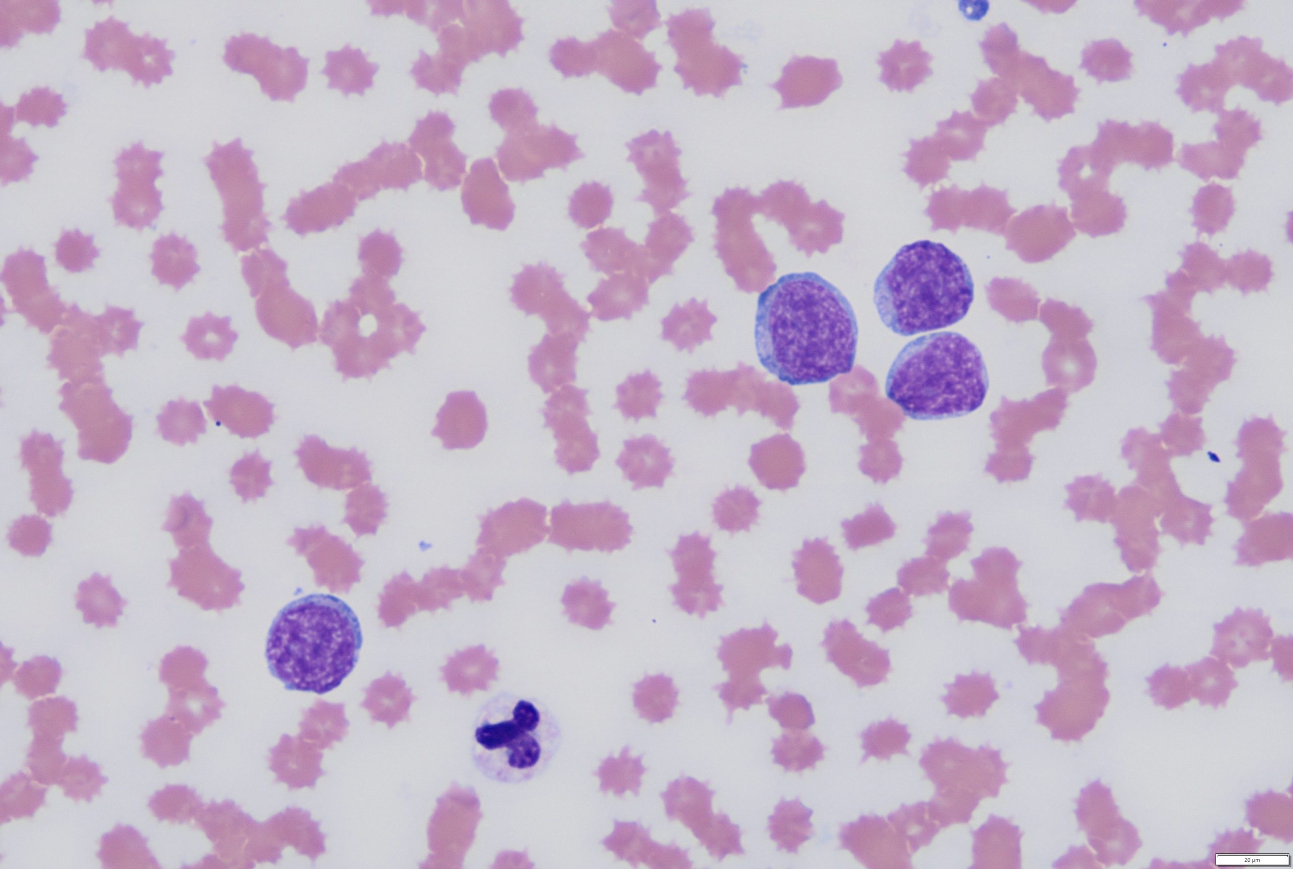
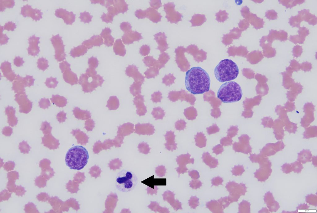
On examination of the blood film, the dog had a lymphocytosis with increased numbers of large atypical mononuclear cells most likely of lymphoid in origin (Figure 1). Instead of a neutrophilia being present, the neutrophil count was low with a degenerative left shift and toxic change. The anaemia appeared non-regenerative.
Put together, these findings were highly suspicious for lymphoid neoplasia (lymphoid leukaemia or leukemic stage of lymphoma). The neutropenia may have reflected secondary infection due to immunosuppression or a combination of effects from the neoplastic process (myelopthisis, tissue necrosis etc.).
Discussion:
This case demonstrates how analyser counts can sometimes be misleading. It is likely that the in-clinic analyser misclassified the neoplastic cells as neutrophils, thereby masking the presence of a neutropenia. Haematology analysers can produce very accurate differential cell counts in healthy animals, but are frequently inaccurate when cells with altered morphology are present e.g. band neutrophils, neoplastic cells, nucleated red cells.
Don’t rely on analyser counts alone. Always review blood smears from sick animals and all cases with abnormal results.
Where possible, a blood film should be examined whenever haematology is performed on a sick animal and any time abnormalities are detected on in-clinic analyser results, whether the animal is sick or well. If a smear examination is not performed, keep in mind that you may be missing something important. For more information on blood film examinations see Kathryn Jenkins article in the June 2022 issue of VetScript.
Thank you to Anna Tiernan, Vet Farm & Pet Clinic, Warkworth, for this interesting case and excellent fresh blood films.