
EMMA GULLIVER
Clinical history
A one-year-old male, neutered, domestic shorthair cat was seen by the referring veterinarian for evaluation of ataxia and a head tilt. Routine haematology and biochemistry were within normal limits, and there were no abnormalities detected on skull radiographs. The cat otherwise seemed well in himself, however clinical signs were progressive and there was no response to treatment with clindamycin (Antirobe®), and his owners elected humane euthanasia.
Laboratory findings
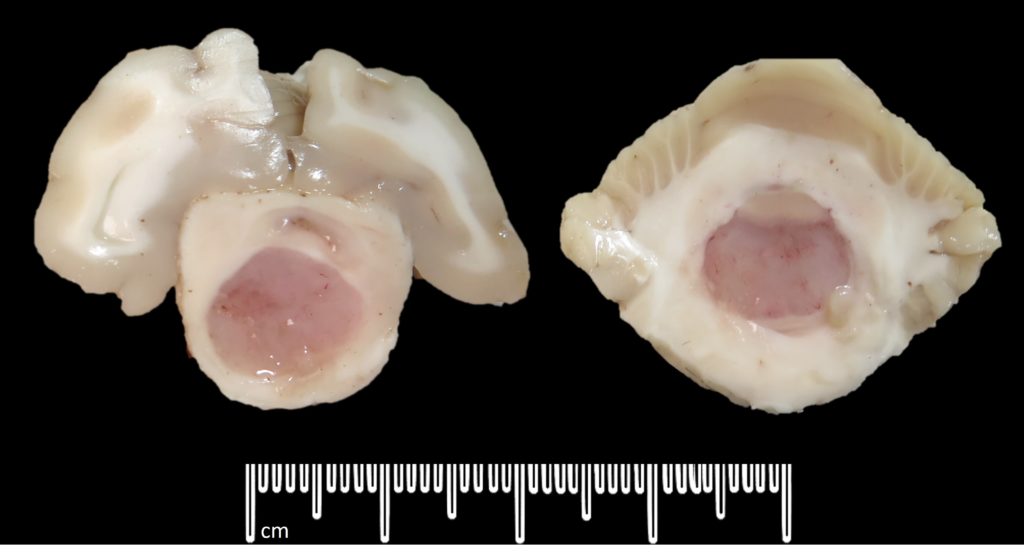
A postmortem examination was performed. There was increased cerebrospinal fluid in the cranium and reddening of the meninges. Serial sectioning of the formalin-fixed brain revealed a pale pink, mottled, gelatinous mass within the brain stem. The mass was slightly right of midline with a maximal diameter of 12 x 11 mm, and extended approximately 3 cm in length from the level of the thalamus rostrally to the level of the caudal cerebellum caudally (figure 1).
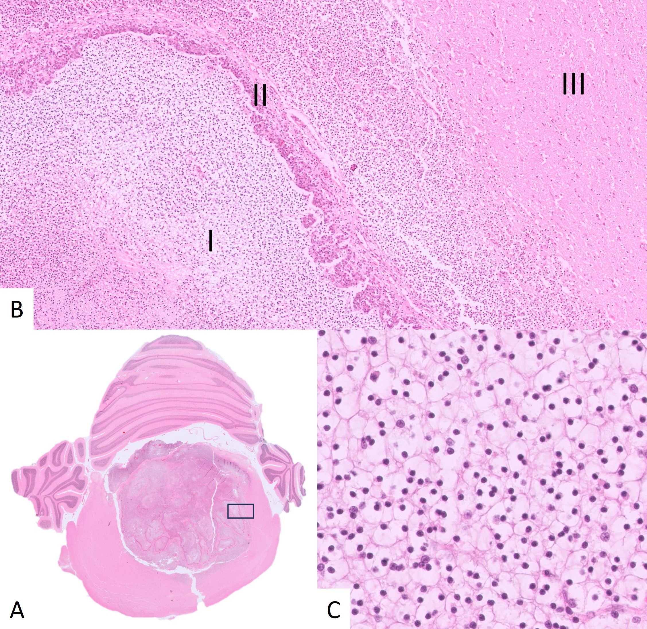
Histopathology (figure 2) revealed a well demarcated, densely cellular and locally infiltrative mass focally expanding and effacing the brainstem. Sheets of polyhedral cells were supported by moderate fibrovascular stroma, often with a prominent glomeruloid-like arrangement. Cells had a small round centrally placed nucleus with condensed chromatin, and abundant clear to pale eosinophilic stippled cytoplasm. Mitoses were not seen.

Laboratory diagnosis
Oligodendroglioma of the brainstem.
Discussion
Oligodendroglioma is a type of glioma arising from oligodendrocytes, the support cells which maintain myelin sheaths around axons in the central nervous system. While it is a relatively common neoplasm in dogs, where it represents up to 15% of primary brain tumours, it is a rare diagnosis in cats with only sporadic case reports in the literature. In both species, meningioma is the most common primary intracranial neoplasm, and is usually seen in aged animals.
The clinical history and young age of this cat initially raised concern for more common differential diagnoses which may result in similar neurological signs. These included bacterial otitis media, central nervous system FIP, bacterial or fungal meningitis, toxoplasmosis or cerebral abscessation. The gross appearance of the mass overlapped with cryptococcosis, which can occasionally form a gelatinous mass lesion in the tissues (a ‘cryptococcoma’), although this would be an unusual presentation.
Gliomas may arise within the grey or white matter of the brain or spinal cord, hence, may lead to a variety of clinical signs which may be sudden in onset and are generally progressive. These may include vestibular signs (head tilt, circling, ataxia), seizures, paraparesis/tetraparesis and changes in behaviour. A clinical diagnosis typically requires advanced imaging (MRI or CT) for identification of a mass lesion. Some gliomas can be diagnosed using intraoperative cytology, although histopathology is more commonly done in veterinary species. There are reports of treatment with surgical excision or radiation, however this diagnosis unfortunately carries a poor prognosis.
Acknowledgements to the owners of this beloved cat and the small animal team at Franklin Vets Pukekohe who submitted this case.
References
> Meuten DJ. Tumors in domestic animals. 5th ed. John Wiley & Sons. 2016.
> Rissi DR, Miller AD. Feline glioma: a retrospective study and review of the literature. Journal of feline medicine and surgery, 19:1307-1314, 2017.