
Clinical history:
A seven-year-old female rabbit was presented for a swelling on the right hind limb, which was worsening after being first noticed two weeks previously. At presentation, the rabbit had muscle atrophy in both hind limbs, with a hairless, nodular mass noted directly above the right calcaneus. The mass was sampled by fine needle aspirate for cytology.
Cytology findings:
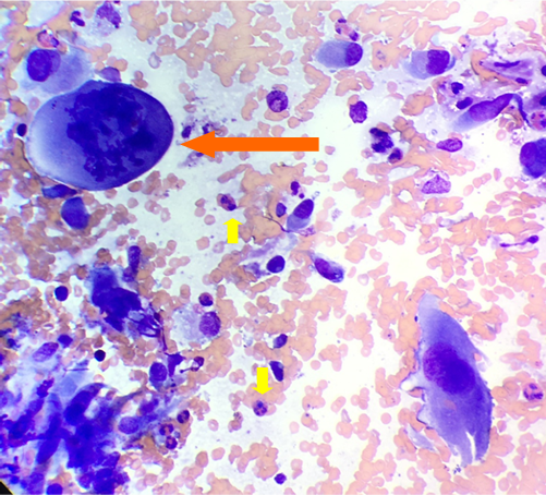
The smears had high cellularity comprising numerous highly pleomorphic spindle cells, amidst thick blood on a proteinaceous background. The spindle cells had marked amounts of basophilic finely granular cytoplasm, often with wispy cytoplasmic tails, large oval to irregular nuclei (up to 70μm diameter), with stippled to coarse chromatin and 2-6 prominent nucleoli. The cells displayed marked anisocytosis and anisokaryosis, with macronucleoli, irregular nucleoli and marked anisonucleoliosis. Frequent karyomegaly with binucleate and multinucleate cells noted (up to 10 nuclei), often of varying nuclear size, including satellite (micro) nuclei. Frequent mitotic figures noted, often large and atypical with lag chromosomes (see Figure 1). Occasional clumps of smaller shrunken cells were also noted, with hypereosinophilic cytoplasm and pyknotic nuclei (necrosis). There were increased heterophils and occasional macrophages, with no infectious agents identified.
Diagnosis:
Anaplastic sarcoma with giant cells.
Discussion:
When multinucleated giant cells are seen in cytologic preparations, differentials include granulomatous lesions (e.g. macrophages in mycobacterial infections), bone lesions (osteoclasts) and neoplastic spindle cells. The marked nuclear atypia, especially variably sized nuclei and satellite nuclei within the multinucleated cells, supported a neoplastic origin in this case.
A recent report on 90 giant cell soft tissue sarcomas from domestic rabbits, divided the cases into two tumour entities, anaplastic sarcoma with giant cells, and histiocytic sarcoma.
Anaplastic sarcoma with giant cells was more commonly reported (72/90 cases). Similar to the reported case, the majority of tumours (86%), were present in the dermis or subcutis, with legs being the most common location (40%). Other reported locations included thoracic and abdominal wall, head and inguinal region. Distant metastasis to lungs, kidney, liver and heart were reported in a low number of cases. CD204 was negative in these cells.
Histiocytic sarcoma was reported in fewer cases (17/90), and most frequently occurred in the lungs, with a quarter of rabbits having a concurrent pneumothorax (likely secondary to tumour effacement of lung tissue). Other reported locations included mediastinum, liver, kidney, cardiac and skeletal muscle tissue. Multinucleate giant cells can be massive, with up to 300 nuclei reported in some cases. These cases stained positive for CD204 via immunohistochemistry (supporting histiocytic lineage).
Both entities were observed in older rabbits (median 7 to 8 years-old). They both appear highly cellular on histopathology, with frequent multinucleated cells and atypical mitotic figures, with large areas of necrosis, haemorrhage and mixed inflammation. Both entities appear highly invasive with strong criteria of malignancy, with evidence for metastatic spread in some cases. For cases occurring on the leg, complete excision is difficult, and high recurrence rate is likely. Overall prognosis is considered guarded.
Thanks to Wainuiomata Carevets for this interesting case.
Reference:
Bertram CA, Garner MM, Reavill G, Klopfleisch R, Kiupel M. Giant cell sarcomas in Domestic rabbits (Oryctolagus cuniculus). Veterinary Pathology. 57:490-496, 2020