MICHAEL HARDCASTLE
Clinical history:
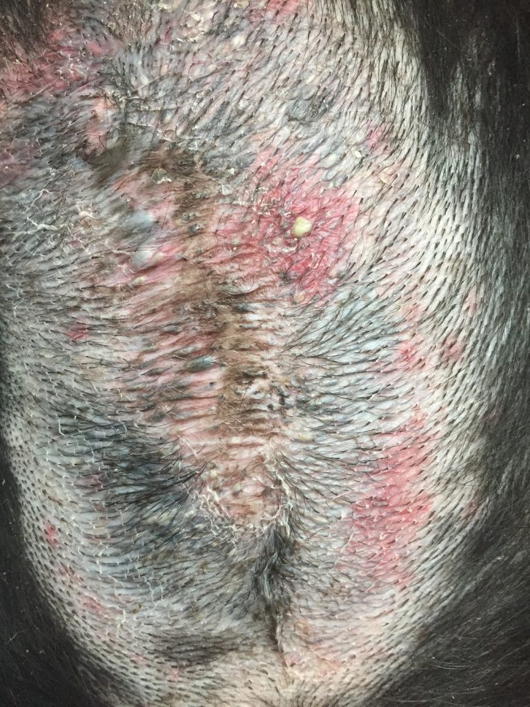
A three-year-old Collie cross had a two and a half year history of pruritic skin and ear disease. This included a dominant sternal rash that had been treated with a range of systemic medications, topical medications and dietary manipulation, but had never resolved; it crusted or flared intermittently. At presentation there was a large area of alopecia, patchy erythema and skin thinning with pustules and comedones on the sternum (Figure 1).
Laboratory testing:
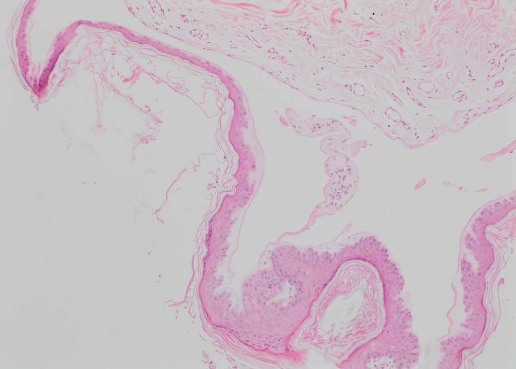
The area was biopsied and histopathology revealed changes of epidermal and follicular hyperplasia with hyperkeratosis, comedones, dermatitis, folliculitis and an oedema-like space in the superficial dermis (Figure 2).
Diagnosis:
Topical corticosteroid reaction
Discussion:
This under-diagnosed syndrome is caused by localised use of potent topical corticosteroids, leading to comedones and skin fragility. The inflammation found in these lesions could be partly a result of skin fragility, and partly due to an underlying allergic skin disease or pyoderma; a perpetuating cycle of topical treatment and worsening signs can develop.
It is typically reported on the thinly haired skin of the ventral abdomen of dogs, and so lesions isolated to this site should raise clinical suspicion for the disease. Histologically, its most distinctive feature is superficial collagen homogenisation and attenuation that can lead to clefting of the dermis.
THANK YOU TO DUNCAN GRAHAM OF ANIMAL DERMATOLOGY NZ FOR THIS INTERESTING CASE.