
KATHRYN JENKINS
On blood smear evaluation and in cytology, eosinophils have distinctive morphological features that make them easy to identify. However, this is not always the case. Here are some surprising tips about the humble eosinophil, and a case report when all was not as it seemed on the haematology analyser results.
Typical eosinophils
Eosinophil granules have an affinity for eosin (the red dye in our routine stains), so that after staining the granules appear pink to orange-red in most species. However the shape, size and number of granules varies considerably between species. The cytoplasm of eosinophils is pale blue (in contrast to other granulocytes), and the nucleus appears less lobulated than neutrophils.
Many species have round granules, however domestic cats have rod shaped ones (Figure 1a). Dogs can have a large variation in size of granules and often display a few cytoplasmic vacuoles (Figure 1b). Granules in ruminant eosinophils are small (Figure 1c). Equine granules are very large, and in this species eosinophils are like finding ‘raspberries’ down the microscope (Figure 1d).
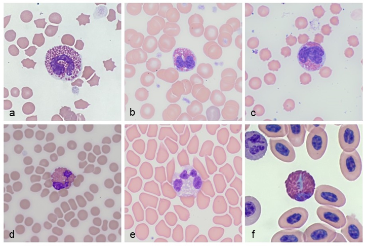
Figure 1. Eosinophil appearance in different species. Cat (a), dog (b), sheep (c), horse (d), greyhound (e), takahe (f).
Grey eosinophils
Eosinophils can however be tricky. In Greyhounds and other sighthounds (and occasionally in other dog breeds), we can see so called ‘grey eosinophils’. In these cases the granules are poor to non-staining, and the cytoplasm often appears highly vacuolated (Figure 1e). This is especially apparent in quick stained smears (like Diff-Quik).
It is important to recognise grey eosinophils, as they can be mistaken for toxic neutrophils or vacuolated monocytes which may suggest an inflammatory process. Grey eosinophils have also recently been reported in cats.
Blue eosinophils
Anyone examining avian blood films may appreciate the wide variety of leukocyte morphology across the many species we see in New Zealand. Heterophils have red-orange elongated granules, and eosinophils can vary from having orange-pink granules to blue granules.
Blue eosinophils can be seen in some psittacine species (especially parrots and cockatoos), as well as our own Kakapo and Takahe (Figure 1f). To complicate the matter, toxic change in heterophils often results in the presence of blue to purple granules and blue cytoplasm, which can mimic blue eosinophils.
Analyser error
A recent case highlighted the value of blood film evaluation. A 4-year-old cat presented with inappetence, weight loss, and fever. On physical exam an enlarged kidney was palpated. Routine biochemistry demonstrated mild hypercalcemia (3.02; ref 2.22-2.67 mmol/L), with mild hypoalbuminemia (26; ref 33-43 g/L). The CBC reported a mild non-regenerative anaemia, with a moderate eosinophilia (7.03; ref 0 – 0.9 x 10⁹/L), and mild monocytosis (3.64; ref 0 – 0.9 x 10⁹/L).
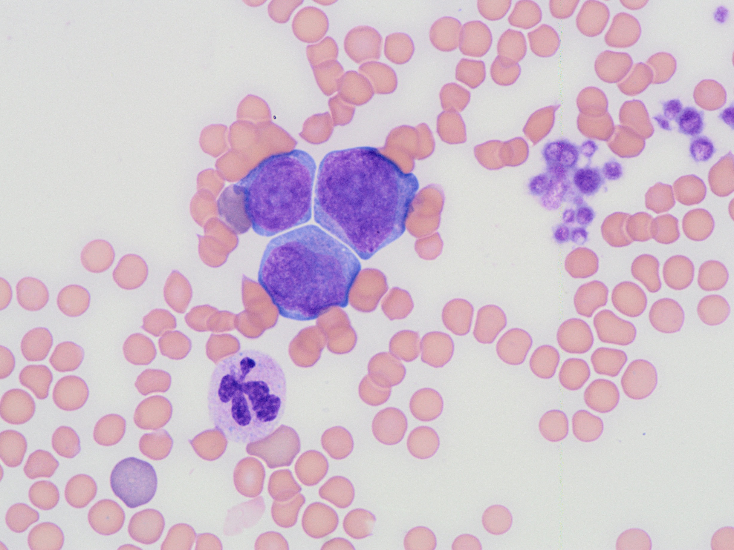
In contrast, on film review, eosinophils and monocytes appeared within normal reference values. There was a significant left shift with toxic change to neutrophils, and moderate numbers of large blast cells with prominent nucleoli (see Figure 2). The interpretation was supportive of lymphoproliferative disease (likely stage V lymphoma), with concurrent systemic inflammation. Subsequent cytology of the kidney was consistent with renal lymphoma.

Figure 2. Cluster of three large ‘blast’-like cells with prominent nucleoli.
In this case, the analyser likely misidentified the inflammatory neutrophils as eosinophils, and the neoplastic blast cells as monocytes. Many haematology analysers use cell size and complexity to categorise cell type, which can differentiate leukocytes extremely well in healthy patients. However when atypical leukocytes are present, the analyser will place the cell into a category of best fit, and will usually “flag” the result. This means a blood film review is critical to check the automated data.
Ideally all blood films should be reviewed to identify atypical cells, and especially when an analyser flags a result, in unwell patients, or the results do not fit with what is clinically suspected.
References
> Stacy NI, Ackerman SJ. A tribute to eosinophils from a comparative and evolutionary perspective. J Allergy Clin Immunol. 147:1115-1116, 2021;
> Holmes, E, Raskin R, McGill P, Szladovits B. Morphologic, cytochemical, and ultrastructural features of gray eosinophils in nine cats. Vet Clin Pathol. 50:52-56, 2021;
> Veterinary Hematology – A Diagnostic Guide and Color Atlas. Harvey, J. 2012.