
KATHRYN JENKINS
A recent interesting case highlighted the issue of a lesion which can mimic the clinical appearance of a benign lipoma.
Clinical history:
A 10-year-old dog presented with a 7 cm diameter mass located on the upper forelimb, which had been slowly growing over the last 4-6 months. The mass palpated like a lipoma, and greasy material was noted when making the smears for cytology. Four smears were submitted for pathologist review.
Cytological findings:
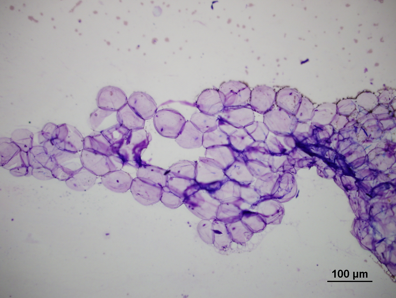
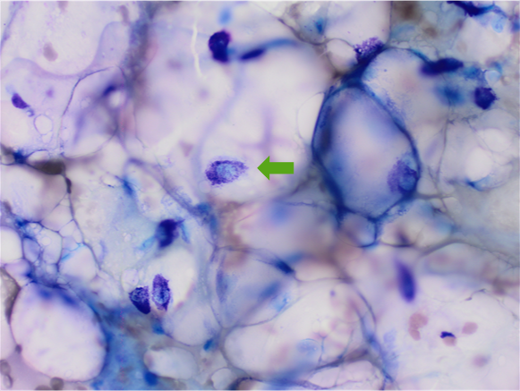
Cell preservation was excellent and the cytology comprised several dense clumps of mesenchymal tissue embedded within a large amount of fat. The spindle cells appearing variably vacuolated with mild to moderate nuclear pleomorphism (Figure 1). Given the unusually large amount of stromal tissue, surgical biopsy for histopathology was recommended. This was performed and confirmed a well-differentiated low grade liposarcoma.
Discussion:
Liposarcoma is the rare malignant counterpart to a benign lipoma. Liposarcomas can vary in appearance histologically (from well-differentiated, anaplastic, or myxoid variants). And as a result they can also vary in clinical appearance, from mimicking lipomas as in this case, through to presenting as a firm infiltrative mass. Although these tumours are considered rare in veterinary medicine, Shetland sheepdogs may be predisposed. Similar to other soft tissue sarcomas, liposarcoma are locally infiltrative, with distant metastasis less commonly reported.
In contrast, lipomas are a common benign tumour of well-differentiated adipocytes in dogs. These are less commonly found in other species, although Siamese cats may be over-represented. Lipomas are often well circumscribed, unencapsulated, freely moveable soft masses, found most often on the trunk and proximal limbs. They are slow growing expansile masses, often cured by complete excision. Grossly the aspiration of fat appears clear and glistening, with a slide that does not dry completely. On cytology the adipocytes of lipomas are indistinguishable from normal fat, and appear as large round balloon-like cells, with a compressed nucleus to the side of the cell. The cohesive adipocytes often have a characteristic “chicken wire” type appearance on low power (Figure 2).

In addition, a small percentage of lipomas occur as infiltrative forms. These tumours invade adjacent connective tissue and skeletal muscle, appearing marbled in appearance grossly. They are also considered benign in behaviour, however they can be more difficult to completely excise.
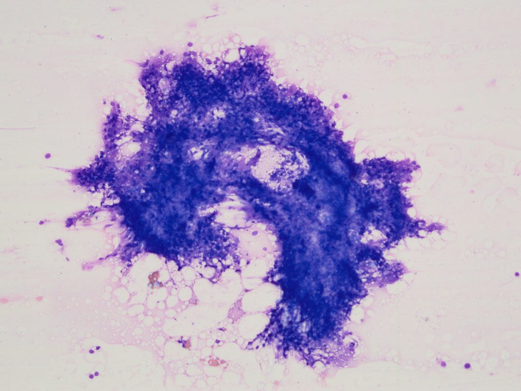
Although liposarcoma are considered rare, other types of soft tissue sarcoma (especially perivascular wall tumours), and subcutaneous mast cell tumours, can also mimic a benign lipoma on palpation, and produce fatty material on cytology slides (Figure 3).
In these cases the power of cytology to provide a useful interpretation is improved by sampling multiple areas within the same mass.
Thank you to David Sheehan, CareVets Kilbirnie for this interesting case.