

BERNIE VAATSTRA
Clinical history
An adult pony mare developed chronic progressive weight loss over the course of 9 months. Clinical examination revealed increased respiratory effort and a draining lesion of the right mandible with bony proliferation. Ultrasound examination and radiography of the lungs revealed multinodular soft tissue opacities throughout the lung fields. Biochemistry and haematology were unremarkable aside from elevated fibrinogen (11 g/L), consistent with inflammation.
The pony was euthanised and post-mortem examination was conducted.
Post-mortem results
The lungs contained numerous nodular fibrous lesions throughout all fields, correlating with the imaging findings. Apart from the mandible lesion noted on clinical exam, there were no other significant gross findings.
Samples of multiple organs were collected into 10% formalin for histopathological examination.
Histopathology
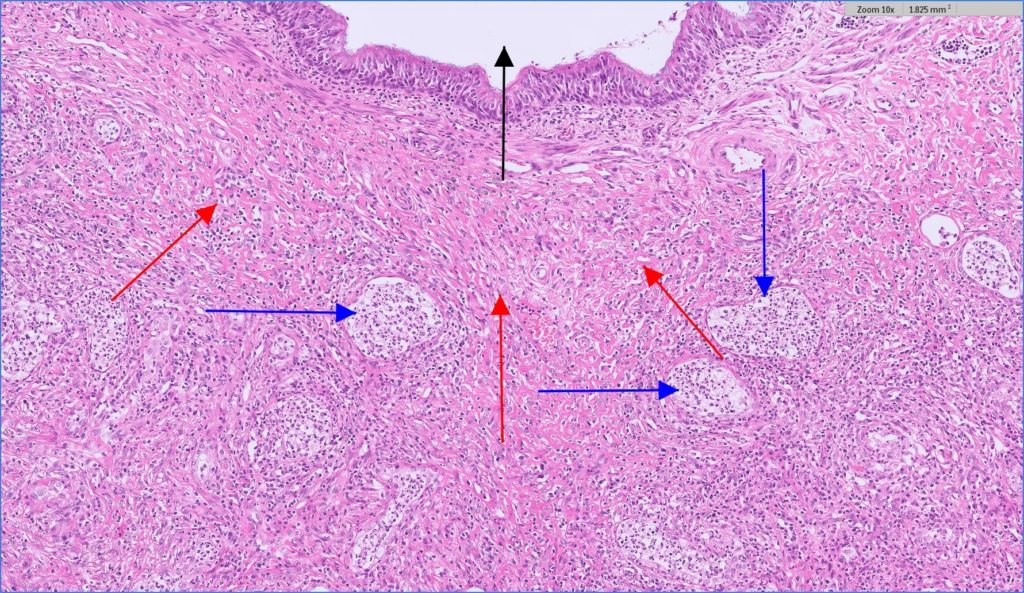
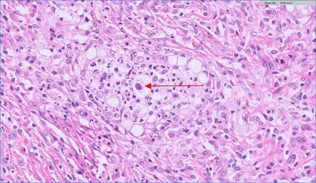
The lung was disrupted by multinodular coalescing areas of fibrosis and consolidation. Airways in affected areas contained aggregates of neutrophils and foamy macrophages. The interstitium was expanded by extensive fibrosis and infiltrated by moderate numbers of neutrophils, macrophages, plasma cells and a few eosinophils, lymphocytes and Mott cells (Figure 1). Alveoli were frequently lined by plump cuboidal cells with hyperchromatic nuclei (type II pneumocyte hyperplasia). Rare macrophages contained deeply eosinophilic intranuclear inclusions that marginated the chromatin (Figure 2). Special stains for bacteria, mycobacteria, and fungal organisms were negative.
The mandible lesion consisted of a focus of bony lysis, fibrosis, and remodelling suggesting a reparative response to an old injury or infection. This was likely not related to the lung lesions.
A sample of tracheal wash fluid collected prior to euthanasia was positive for Equine herpesvirus-5 using real-time PCR.
Incorporating the gross, histological and molecular findings, the final diagnosis was confirmed as equine multinodular pulmonary fibrosis due to EHV-5 infection.
Discussion
Equine multinodular pulmonary fibrosis is a progressive lung disease of horses strongly associated with the presence of EHV-5, a gamma-herpesvirus. Affected horses tend to be middle-aged and older and clinical findings include chronic weight loss, fever, tachypnoea, wheezes and crackles on auscultation. Clinical pathology findings are non-specific but may include increased fibrinogen, leukocytosis and anaemia (Wilkins 2013).
Differential diagnoses include recurrent airway obstruction, infectious pneumonia (fungal, bacterial), foreign body aspiration, and pulmonary neoplasia.
Antemortem diagnostic aids include transtracheal wash cytology to assess for inflammation and look for rare macrophages with intranuclear inclusions, imaging studies to demonstrate the characteristic nodular interstitial pattern, and airway fluid culture and PCR for EHV-5.
It is noteworthy that EHV-5 is highly prevalent in horses and most infections are
asymptomatic. In a small percentage of horses, appropriate host factors such as age and immunological response could favour activation of virus within T and B lymphocytes, leading to clinical disease. EVH-5 has also been associated with lymphoproliferative disease, with rare reports of horses developing EPMF and lymphoma concurrently (Bawa et al, 2014).
The prognosis for equine multinodular pulmonary fibrosis is unfortunately poor in most cases, with one study reporting 14% survival beyond 6 months after diagnosis (Easton-Jones, et al 2019). Treatment with antivirals plus or minus glucocorticoids has been reported to be effective in a small number of cases.
Acknowledgements to Rachael Smith from the Rangiora Vet Centre for submitting this interesting case.
References:
Bawa B, Vander Werf K, Beard L, Davis E, Andrews G, and Almes K. Equine Multinodular Pulmonary Fibrosis and Lymphoma in a Horse Associated with Equine Herpesvirus-5. Journal of Equine Veterinary Science, 34:694-700, 2014.
Easton-Jones CA, Cissell DD, Mohr FC, Chigerw M, and Pusterla N. Prognostic indicators and long-term survival in 14 horses with equine multinodular pulmonary fibrosis. Equine Veterinary Education, 32:41-6, 2020.
Wilkins PA. Equine multinodular pulmonary fibrosis: Diagnosis and treatment. Equine Veterinary Education, 25:393-6, 2013.