
SANDY WELTAN
The cat or dog that ain’t doing right. We all see those frustrating cases, slight lethargy, intermittent vomiting, abdominal pain, and non-specific abnormalities in biochemistry or haematology. Some may present acutely ill with jaundice and elevated liver enzymes. Tests for pancreatitis may or may not be positive and ultrasound may identify abnormalities in some cases. Detection of bile sludge could be an indication for aspiration of bile (Verwey et al 2023).
Cytology is the most useful tool for detecting cholecystitis in dogs and cats (Peters et al 2016). Aspiration of bile is easily achieved with the aid of ultrasound with rare complications.
Three cases of cholecystitis are described.
Case 1
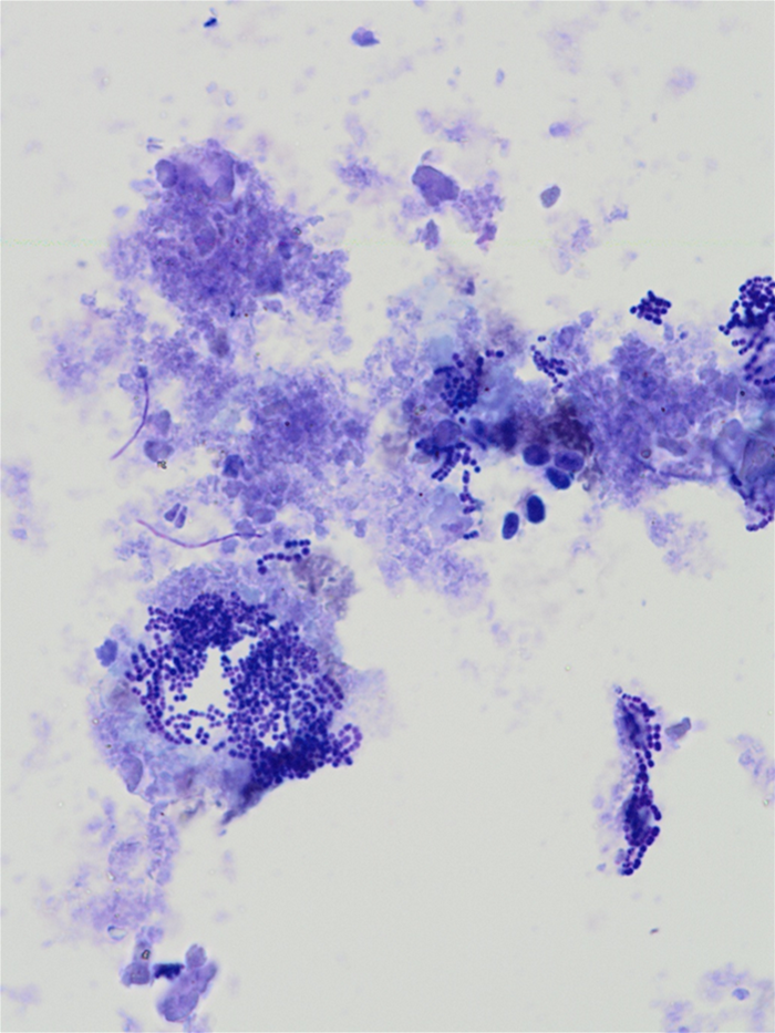
“Joshua”, a 14-year-old domestic short-haired, male cat presented with persistent jaundice but generally well with intermittent vomiting. Bloods and ultrasound did not narrow down the cause. Medication for hepatic support had no effect. On exploratory laparotomy, the gall bladder was found to be flocculent and floppy but very inflamed and vascular. A sample of bile was submitted to the laboratory (figure 1).
Bacteria and yeasts are present but the only organism that grew on culture was a non-haemolytic Streptococcus sp..
Case 2
“Max” 14-year-old male Schnoodle, only eating treats or hand-fed for four to five days. No vomiting or diarrhoea, normal activity. In house biochemistry, elevation 22x ALT, 14x ALP, three days later elevated bilirubin and cholesterol.
Urine and serum samples were submitted for leptospirosis PCR, liver and bile aspirates for cytology. The liver cytology was non-specific (figure 2). Culture yielded heavy growth of Enterococcus caecorum.
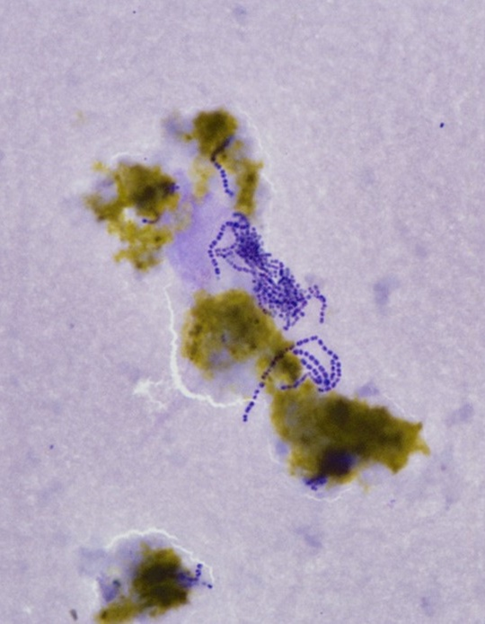
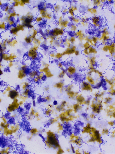
Figure 2a and 2b. Bile cytology from case 2. Culture yielded heavy growth of Enterococcus caecorum.
Case 3
“Spunky”, 13-year-old male DSH cat. Unwell, anorectic, ALT >1000, cystic lesions present in liver.
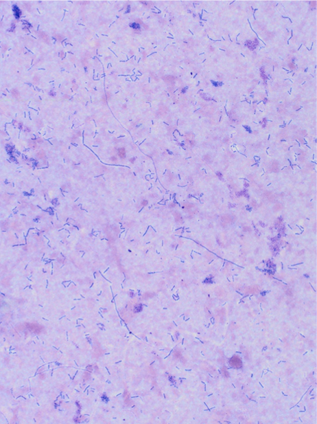
Green fluid was submitted to the laboratory. Direct and cytocentifuged smears were made and examined (figure 3).
Culture yielded heavy growth of Citrobacter braakii. Histopathology was not performed but the multiple cysts, some containing bile and some clear fluid could suggest Caroli’s disease secondary to ductal plate malformation.
Discussion
Infection may also be associated with choleliths and mucocoeles. Both cytology and culture are recommended on bile for diagnosis of cholecystitis (Peters et al 2016, Pashmakova et al 2017). Small numbers of bacteria have been detected in normal dogs on cytological examination (Verwey et al 2021). Neutrophils often do not survive in the hostile environment of bile so the presence of intracellular bacteria cannot be used to confirm infection. There are also microorganisms that may be detected on cytology that may not grow on culture, notably fungi and anaerobic bacteria (Verwey et al 2023).
References
Pashmakova MB, Piccione J, Bishop MA, Nelson WR, Lawhon SD. Agreement between microscopic examination and bacterial culture of bile samples for detection of bactibilia in dogs and cats with hepatobiliary disease. J Am Vet Med Assoc 250:1007-1013. 2017.
Peters LM, Glanemann B, Garden OA, Szladovits B. Cytological Findings of 140 Bile Samples from Dogs and Cats and Associated Clinical Pathological Data. J Vet Intern Med 30:123-131. 2016.
Verwey E, Gal A, Kettner F, Botha WJ, Pazzi P. Prevalence of subclinical bactibilia in apparently healthy shelter dogs. J Small Anim Pract 62:948-958. 2021.
Verwey E, Weltan SM, Whitehead Z. Candida albicans cholecystitis in a dog with diabetes mellitus. Vet Rec Case Reports 11:e630. 2023.