
Arefeh Ravanbakhsh
Pelger-Huët anomaly (PHA) is a genetic condition reported in numerous species characterised by nuclei hyposegmentation of granulocytes (neutrophils, eosinophils, and basophils).
Nuclei of granulocytes can appear oval, kidney-shaped, bean-shaped, band-shaped or bilobed.1 This anomaly has been described in dogs, cats, horses, rabbits, and humans.1 It is thought that a defect occurs at the stem cell level as bone marrow megakaryocytes can also be affected.2 In humans. Pelger-Huët anomaly has been attributed to a mutation in lamin-B receptor gene which codes an integral nuclear envelope protein.2 In most cases, this is an autosomal dominant hereditary anomaly, with no clinical consequences in heterozygous individuals; however, it can be lethal in homozygous individuals.2,3
Although uncommon, it is important to be aware of this anomaly to avoid misinterpretation of acute inflammation when examining blood smears.
Assessment of a blood smear as part of a routine CBC (such as a presurgical screen) in an otherwise healthy patient can be a fantastic way to pick up on this anomaly. In patients with PHA almost all of the neutrophils are hyposegmented. At first glance it may appear that the patient has a severe left shift, suggesting an overwhelming inflammatory response. However closer scrutiny of the neutrophils and other granulocytes reveals several clues to differentiate PHA from acute inflammation.
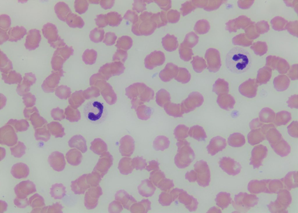
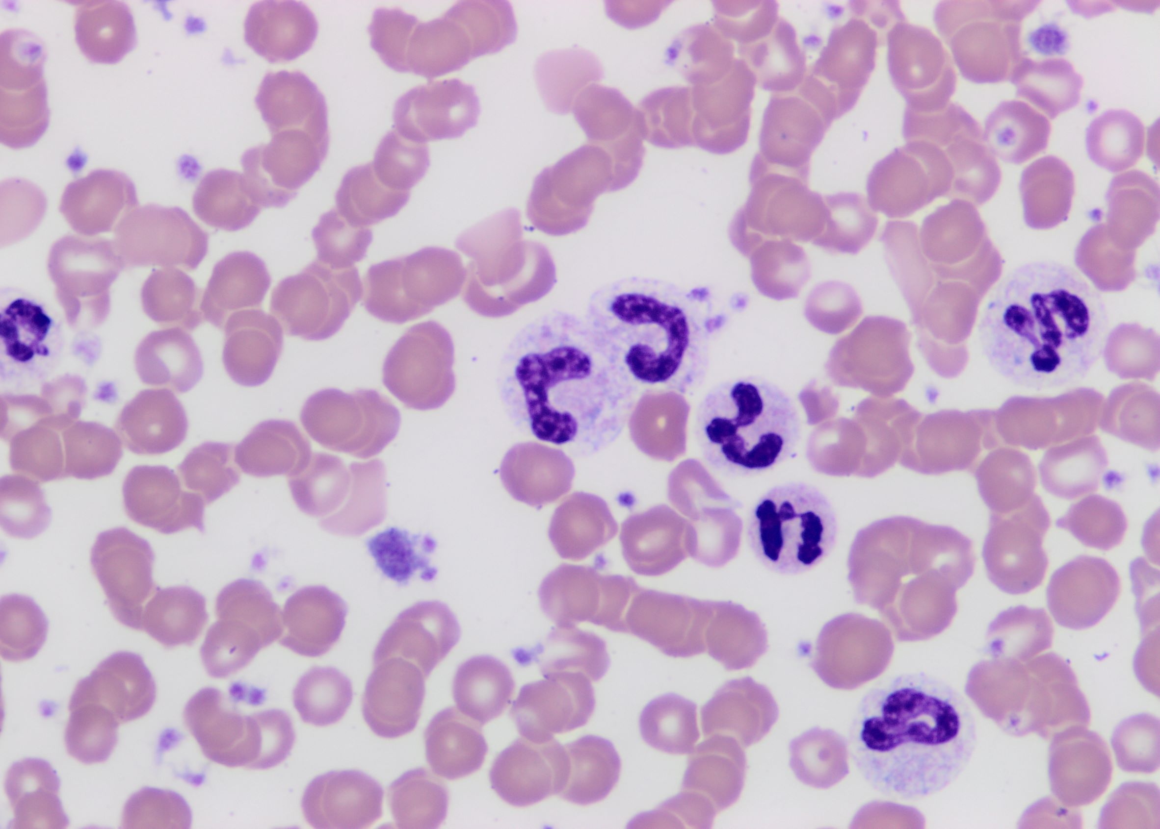
Figure 1 shows a blood smear from a patient with PHA while Figure 2 is from a diabetic patient with evidence of acute inflammation.
What differences can be appreciated and how can band neutrophils
be distinguished from segmented neutrophils?
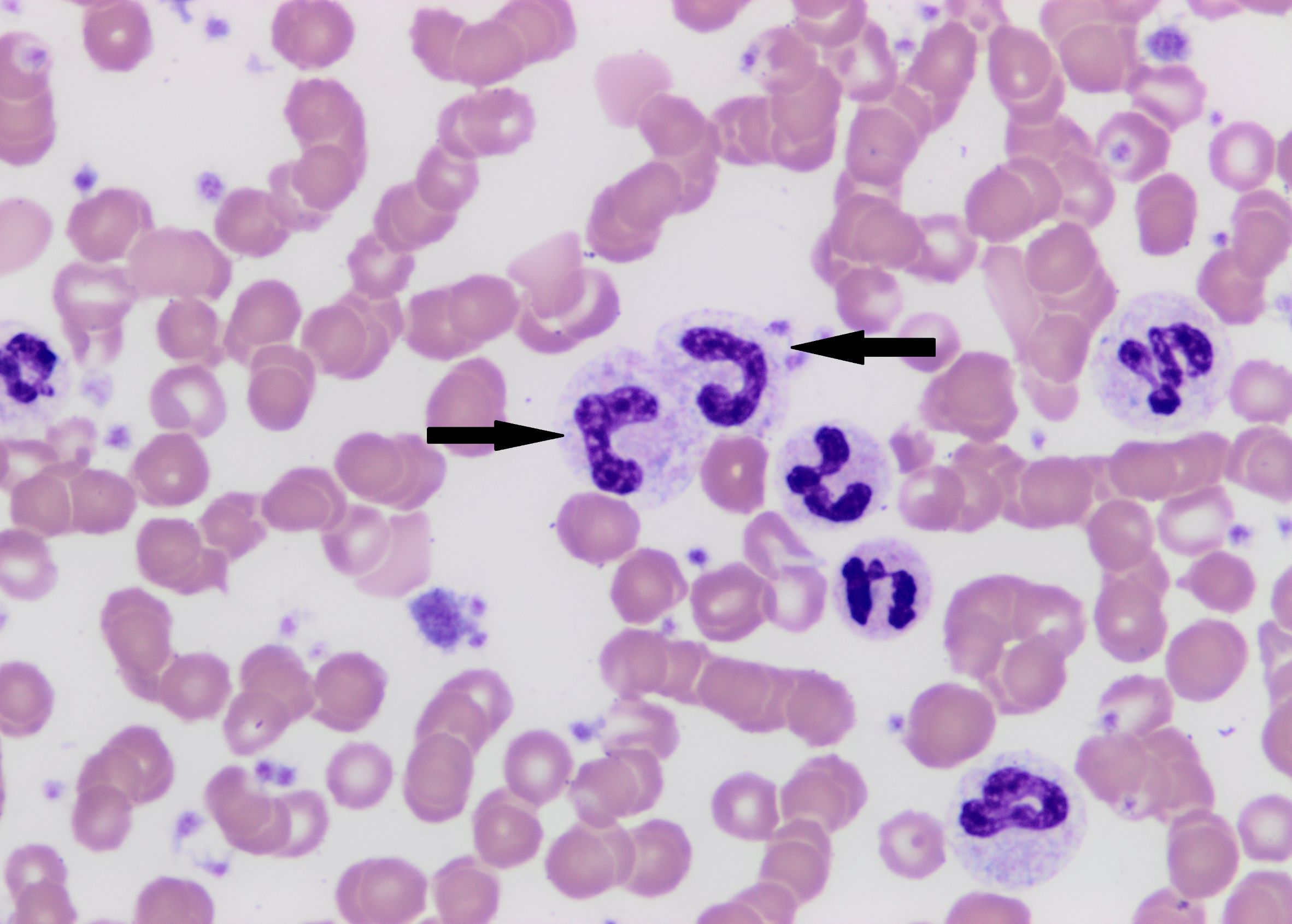
A band versus segmented neutrophil can be differentiated by closely examining the width of most narrow portion of the nucleus and comparing that to the widest part of the nucleus. Segmented neutrophils have a tight constriction between nuclear lobes. The narrowest part of the constriction should be <1/3 the width of the thickest part of the nucleus. If the width of a constricted part of the nucleus is >1/3 the width of the thickest part of the nucleus, the cell is a band. In both Figure 1 and Figure 2 neutrophils with a band-shaped nuclei are present (arrows). Further assessment of their cytoplasmic features and chromatin pattern can aid to determine if the band neutrophils are due to release of immature neutrophils secondary to a strong inflammatory stimulus vs a genetic anomaly.
The cytoplasm of the band neutrophils in Figure 2 display cytoplasmic basophilia and some contain Döhle bodies. Cytoplasmic basophilia, Döhle bodies as well as cytoplasmic vacuolation and toxic granulation are all signs of toxic change which indicates accelerated maturation through the bone marrow supporting an inflammatory response. The banded neutrophils in Figure 1 do not display toxic change and have cytoplasmic features of normal mature segmented neutrophils.
The second clue is the chromatin pattern. The banded cells in Figure 1 have densely clumped chromatin pattern while the banded cells in Figure 2 have more loosely clumped chromatin pattern. Furthermore, patients with the inflammatory response have a mixture of bands and segmented neutrophils. Patients with PHA have uniformly hyposegmented neutrophils.
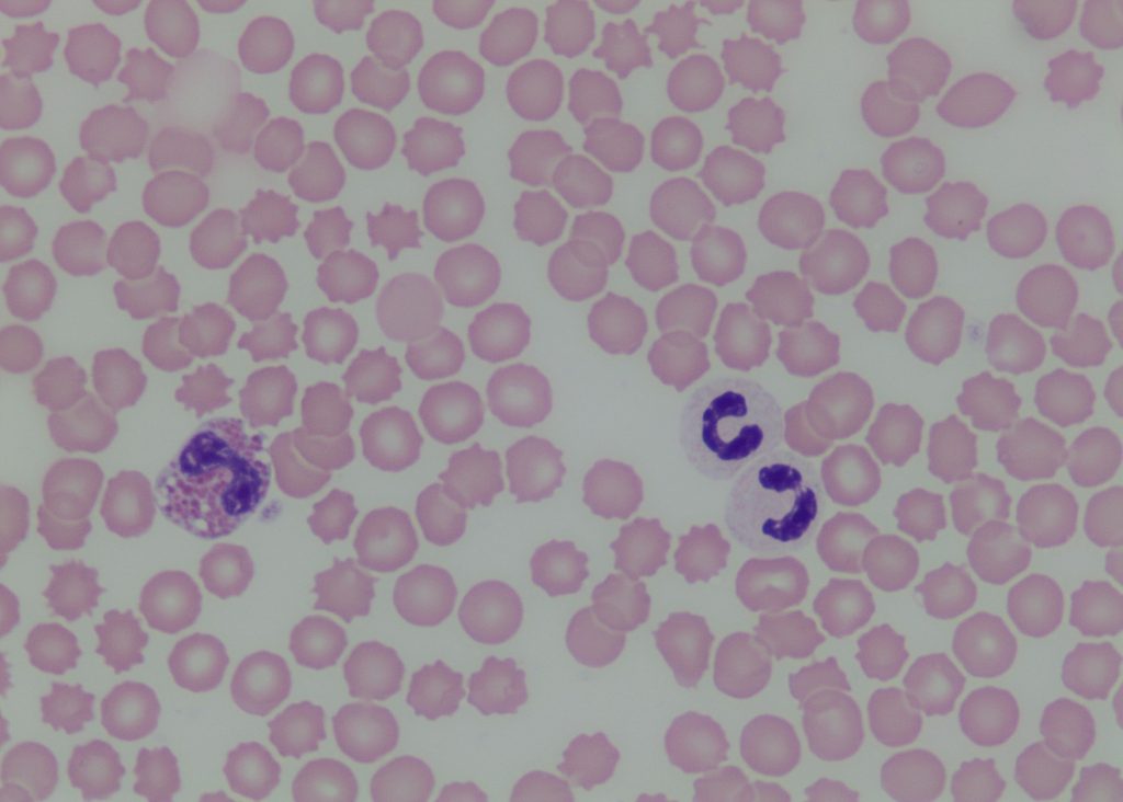
Another helpful hint would be to assess the nuclear morphology of other granulocytes. Figure 3 is from the same patient with PHA. Note that the eosinophil nuclei is also hyposegmented.
Pelger-Huët anomaly should also be differentiated from pseudo-Pelger-Huët anomaly where granulocyte hyposegmentation occurs not due to a genetic condition but rather secondary to other underlying pathology such as myelodysplastic syndrome, chronic infection, or administration of some drugs. In pseudo Pelger-Huët, only a minority of granulocytes display hyposegmentation.2
References:
1. Harvey JW. Veterinary Haematology, A Diagnostic Guide and Color Atlas. Elsevier St. Louis Missouri, USA, 2012.
2. Valenciano AC, Cowell RL. Cowell and Tyler’s Diagnostic cytology and Hematology of the Dog and Cat. 5th Edtn. Elsevier St. Louis Missouri, USA, 2019
3. Vale A.M et al. Pelger-Huët anomaly in two related mixed-breed dogs. J Vet Diagn Invest. 4:863-5, 2011
4. Deshuillers. P, Raskin Rm Messick J. Pelger-Huët anomaly in a cat. Vet Clin Pathol. 3:337-41, 2014.