
JiYe Ahn
History
A 3-year-old female New Zealand Heading dog that had arrived at a farm three weeks previously was found dead in a pool of blood originating from the anus. No external evidence of trauma was identified. On post-mortem examination, haemorrhage of the large intestine was noted. Samples of liver, kidney, spleen, and small and large intestine were collected and submitted to Awanui Laboratory for histopathology.
Histopathology
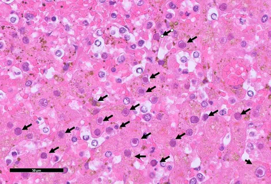
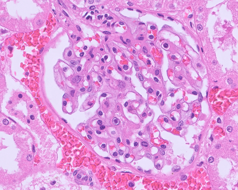
Throughout the entire hepatic lobe, there was necrosis of the centrilobular hepatocytes, occasionally extending into midzonal areas. This was characterized by disruption of hepatic cord architecture and loss of hepatocytes, with replacement by abundant fibrin, haemorrhage, and necrotic debris admixed with low numbers of lymphocytes and plasma cells. Hepatocytes at the periphery of necrotic areas often contained basophilic, 4–5 µm diameter, round to oval intranuclear viral inclusion bodies that were occasionally surrounded by a clear halo and marginate the chromatin. Throughout the hepatic parenchyma, there were numerous foamy Kupffer cells containing intracytoplasmic golden-brown pigment (presumptive lipofuscin, hemosiderin or bile) and the previously described intranuclear viral inclusions. Predominantly centrilobular hepatocytes and the bile duct epithelium contained abundant intracytoplasmic golden-brown pigment (presumptive lipofuscin, hemosiderin or bile). Multifocally within the bile canaliculi were bile plugs. Additionally, intranuclear viral inclusions were identified in renal glomeruli endothelial cells. Other findings included haemorrhage and congestion of the small and large intestines, and diffuse splenic lymphoid depletion.
Diagnosis
The lesions were consistent with infectious canine hepatitis (ICH).
Discussion
Infectious canine hepatitis, also discussed in the September 2020 issue of VetScript, is an “old” disease that occurs sporadically worldwide. Infectious canine hepatitis is caused by canine adenovirus type 1 (CAV-1), which results in viral hepatitis and endothelial damage in canids, leading to haemorrhage. Transmission occurs through direct or indirect contact with contaminated fomites, nasal and conjunctival secretions, faeces, or urine. Most cases present with only a mild febrile illness, such as pharyngitis and tonsillitis. However, severe cases may present with vomiting, melena, high fever, and/or nonspecific neurological signs. Diarrhoea may contain frank blood or melena. Along with haematuria, widespread petechial and ecchymotic haemorrhages—so-called “paintbrush lesions”—may be observed. Corneal oedema (“blue eye”) can occur as a result of viral replication within corneal endothelial cells and a type III hypersensitivity reaction. The liver may show an enhanced lobular pattern, and fibrin strands may be present on the surface. An oedematous gallbladder may also be observed.
Clinicopathological findings include neutropenia, lymphopenia, and thrombocytopenia in the early stages, followed later by neutrophilia and lymphocytosis. Increased alanine aminotransferase (ALT) and aspartate aminotransferase (AST) activities may be present, along with prolonged bleeding time (BT), activated partial thromboplastin time (APTT), and thrombin time (TT).
In New Zealand, diagnosis can be made by correlating history, clinical signs, clinicopathology results and histopathology as molecular testing is limited. Differential diagnoses include toxins such as xylitol or carprofen, chronic passive congestion, or infection with canine herpesvirus 1 (CaHV-1) or canine parvovirus (CPV).
Infectious canine hepatitis should be suspected in dogs with questionable vaccination histories that present with fever and signs of hepatic, gastrointestinal, or respiratory disease.
References:
Hardcastle, M (2020). Infectious canine hepatitis – it’s still out there. VetScript 38(4), 52-53, 2020
Sykes, J. E. (2013). Infectious canine hepatitis. Canine and feline infectious diseases, 182.
Cullen JM, Stalker MJ. Liver and biliary system. In: Maxie MG, ed. Jubb, Kennedy, and Palmer’s Pathology of Domestic Animals. Vol. 2. 6th ed. St. Louis MO: Elsevier Ltd; 2016:282,310-312.