

LISA HULME-MOIR
We don’t often receive cytology samples from farm animals but just like in dogs and cats, cytology has the potential to yield a quick diagnosis that assists with decision making, optimising animal welfare and promoting judicious use of antibiotics.
Clinical history:
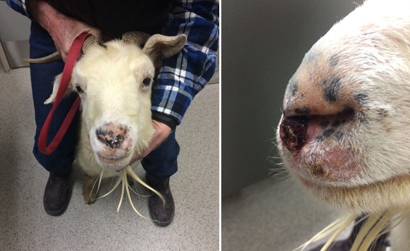
A fine needle aspirate was submitted from a lesion on the nose of a 14-year-old Cashmere doe (Figure 1). The lesion had not been visible 2 weeks previously when the doe had her teeth examined, and it did not appear to be bothering her.
Laboratory testing:
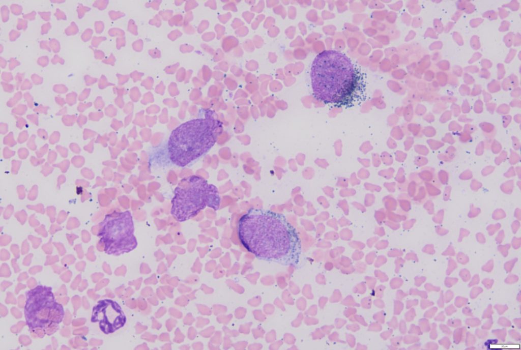
On cytology, multiple large round to spindle-shaped cells containing fine black pigment were present (Figure 2). The cells were moderately pleomorphic with moderate anisocytosis and anisokaryosis, occasional giant macrocytes, binucleate cells and frequent large nucleoli. The pigment was consistent with melanin and a diagnosis of melanoma was made.
Discussion:
Melanomas are common skin tumours in goats, particularly fibre breeds such as Angora, Pygora and Cashmere.1 In one study of Angora goats in Queensland, melanomas were the second most common skin tumour with a prevalence of 2.2%, behind squamous cell carcinomas, which had a prevalence of 3.8%.2 This predisposition has led some to propose Angora goats as a potential model for studying melanomas in humans.
The tumours most commonly occur on the dorsal surface of the ear, although other sparsely haired areas of the body with high UV exposure such as the nose or perineal area are also common locations for melanomas.
In the present case due to the old age of the doe, a palliative approach was taken with the plan to euthanase when the lesion started to impact her welfare.
Many thanks to William Cuttance and the team at VetEnt Te Kuiti for the excellent smears and interesting case.
References:
1. Mavangira et al. Malignant melanoma of the horn base in a Pygora goat. Journal of Veterinary Diagnostic Investigation, 20:104-107, 2008.
2. Green at al. An animal model for human melanoma. Photochemistry and Photobiology, 64:577-580, 1996.