
CRISTINA GANS
Clinical history
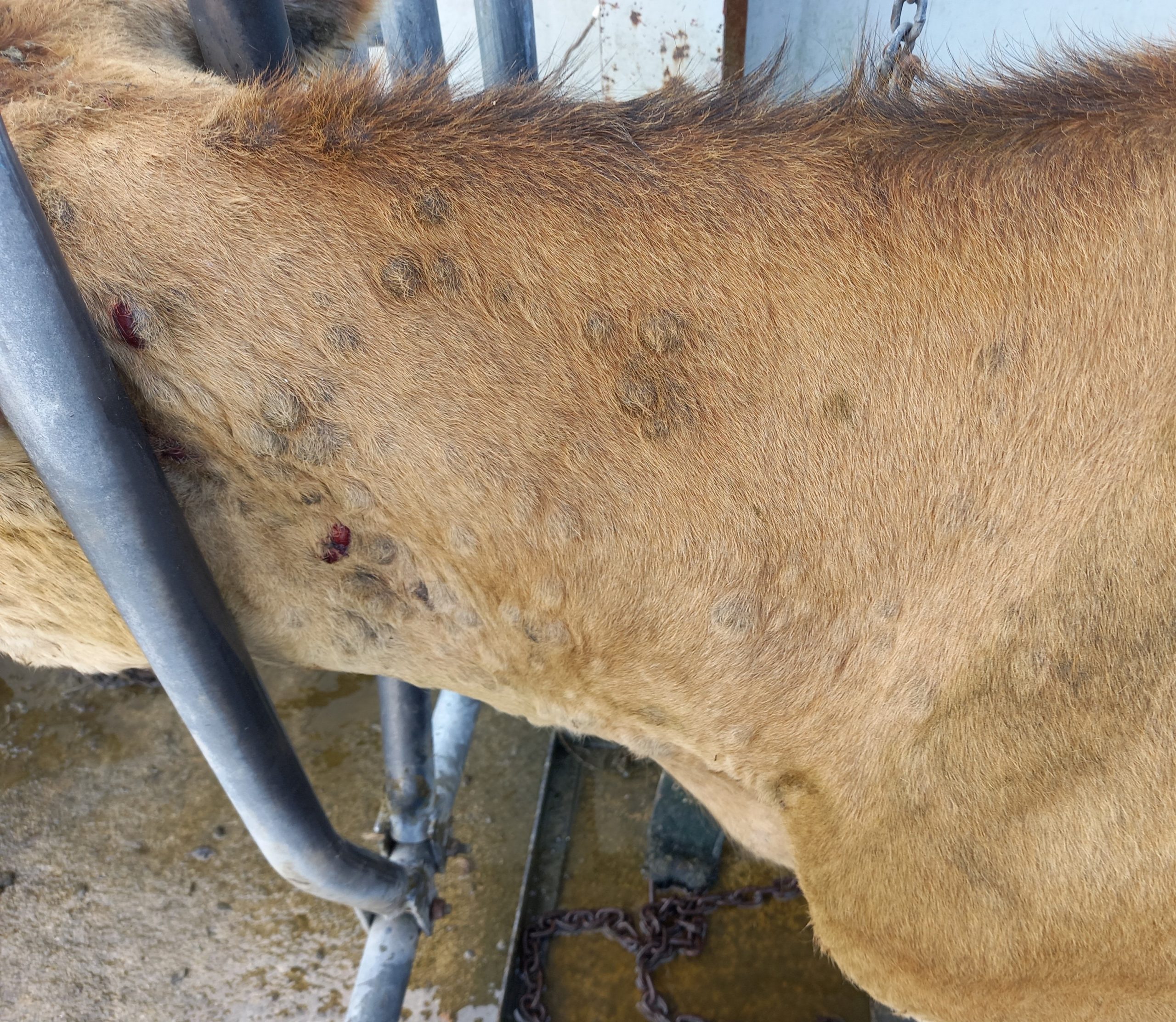
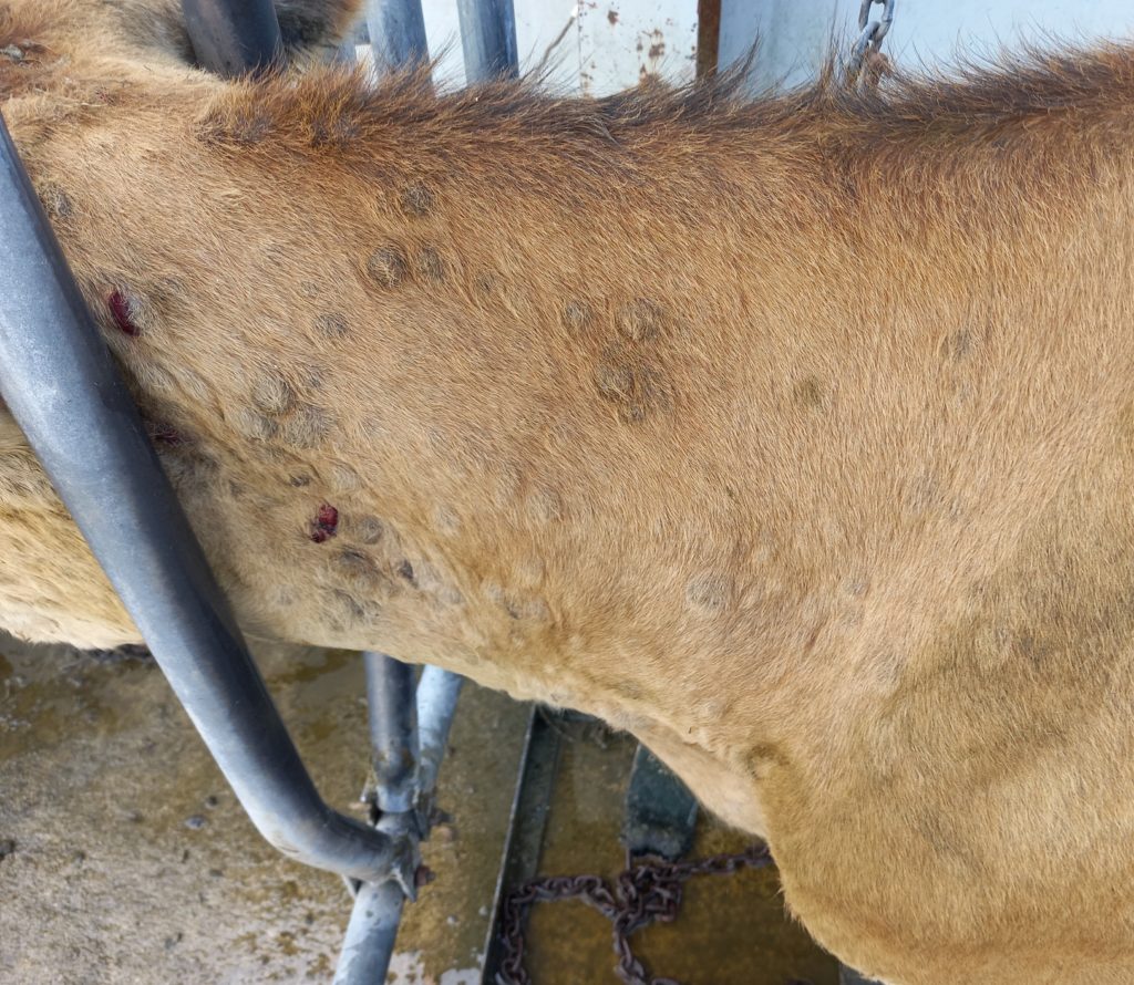
A two-year old Jersey cow presented to the veterinarian with multiple cutaneous nodules of unknown duration, located throughout the body, although most were concentrated on the head and neck (Figure 1). Some nodules appeared ulcerated and may have been secondarily infected. Lymph nodes were not obviously enlarged. Other animals in the herd were not affected. No other significant abnormalities were noted on clinical examination.
Laboratory findings
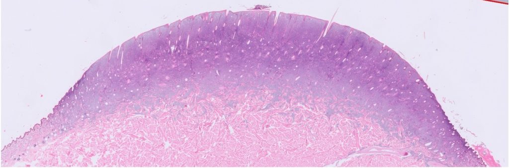
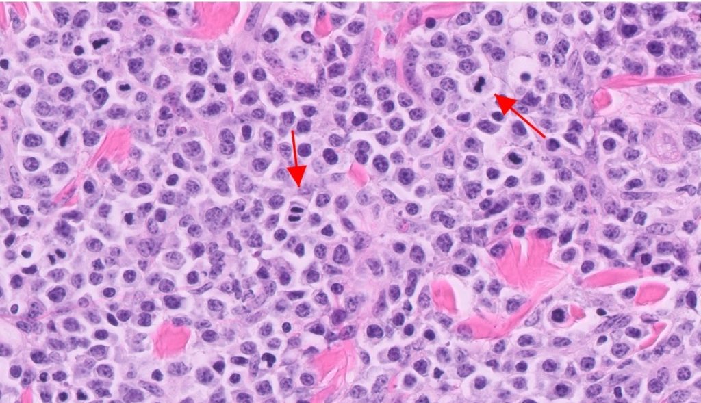
An excisional biopsy of two of the nodules were submitted for histology. Histology of the plaque-like masses revealed a densely cellular neoplasm composed of large lymphocytes infiltrating along the dermis and raising the epidermis (Figure 2). The neoplastic population displayed marked cellular and nuclear atypia. A very high mitotic count of over 200 per 10 hpf (or 20/ hpf) was present (Figure 3).
Laboratory diagnosis
Cutaneous lymphoma
Discussion
Lymphoma is one of the most common reported neoplasms in cattle and is classified as either enzootic or sporadic. Of the sporadic types, at least three different forms of lymphoma in cattle have been described: calf/multicentric, thymic and cutaneous. These forms occur most commonly in animals less than 3 years of age and are not associated with bovine leukaemia virus (BLV).
A rapidly progressing multicentric lymphadenopathy progressing to death characterises the calf form, which in spite of the name, can affect cattle of up to two years of age.
The thymic form is characterised by lymphoproliferative enlargement of the thymus, which depending on the size and location of the tumour, could manifest as a cervical swelling. Other reported clinical signs include dyspnoea, bloat, jugular distention, tachycardia, or oedema of the head and neck.
In comparison to the thymic and calf forms, the cutaneous form presents a less aggressive clinical course, and most typically affects cattle aged 1 to 3 years of age. Lesions may regress and reappear, with eventual progression to visceral organs and the blood. Lymphadenopathy may accompany the skin lesions.
The enzootic form, also known as enzootic bovine leukosis, is associated with BLV infection and is described in adult cattle (with a reported peak incidence 6-8 years of age) Bovine leukosis virus is a oncogenic retrovirus, which infects bovine lymphocytes. Neoplastic lesions may involve the abomasum, heart base, uterus, and other organs, with varied clinical manifestations.
The NZ dairy herd is considered to be free of enzootic bovine leukosis. MPI should be notified in cases of concern for BLV infection, particularly when there is visceral involvement, or a persistent lymphocytosis in an older animal, or if multiple animals in a herd display lesions compatible with lymphoma.
Acknowledgements to Hamish Newton at Veterinary Centre Oamaru for this case submission and photograph.