
JOHN GILL
Malignant catarrhal fever (MCF) is a sporadic disease of cattle and deer, caused by the sheep-associated virus (ovine herpesvirus 2). MCF is passed to cattle and deer by carrier hoggets and lambs, but affected cattle and deer do not transmit MCF to their cohorts. The incubation period can be several months.
The following cases show that this disease can produce a variety of clinical signs, although the “head and eye” presentation involving single adult cattle and acute deaths in two year-old stags are probably the most common.
In the past, this disease was diagnosed by examination of the fixed brain of suspect cases. This has largely been replaced by the use of PCR testing of EDTA blood from live, affected animals or the heart blood of animals found dead.
Case 1
A mature dairy cow was found recumbent and in convulsions. It died shortly afterward. Samples of eye fluid from the dead cow showed normal concentrations of magnesium and calcium.
A necropsy revealed only a large amount of blood in the lumen of the colon.
Histopathological examination of sections of the colon showed severe submucosal oedema, thrombosed arterioles often with large infiltrates of mononuclear cells and necrotic ganglia. The sections of the mucosa revealed necrosis of crypt lining cells and replacement of large areas of the mucosa by sheets of lymphoid cells. These findings were considered to be consistent with MCF of cattle.
Later examination of the fixed brain from this cow confirmed this diagnosis.
Case 2
There was an outbreak of deaths in a group of 275 yearling stud Angus bulls on a sheep and beef farm. These animals were in multiple mobs of 90 animals and most of the affected animal were from one mob.
Approximately eight of these bulls died over a two-week period. Some were found dead, but the majority showed a variety of clinical signs that included a severe oculo-nasal discharge and pyrexia, or recumbency and seizures before dying. All exhibited a peripheral corneal oedema.
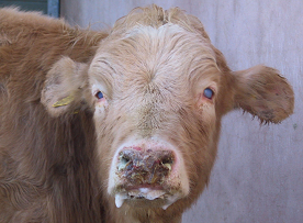
Photo credit: nadis.org.uk
EDTA blood samples from live affected bulls were PCR positive for ovine herpesvirus 2. The fixed brains from a couple of yearlings showed a relatively mild, non-suppurative meningoencephalitis, typical of MCF. As this disease has a long incubation period, it was thought the virus was derived from an adjacent paddock of hoggets that these yearlings spent some months close to over the winter.
Case 3
On a deer farm, ten out of a mob of 100 adult stags were found dead over a 10-day period. A few were noticed to have a bloody diarrhoea shortly before they were found dead. A necropsy of one recently dead stag demonstrated a severe congestion and haemorrhage of the intestinal tract from mid-intestine to the rectum.
Culture of the intestinal tract failed to find a bacterial pathogen, but the ovine herpesvirus-2 was found in EDTA blood taken from this animal, confirming a diagnosis of MCF.
Large outbreaks of MCF were seen in the early days of deer farming in New Zealand, but are rarely seen on today’s deer farms.