SANDY WELTAN
Clinical history:
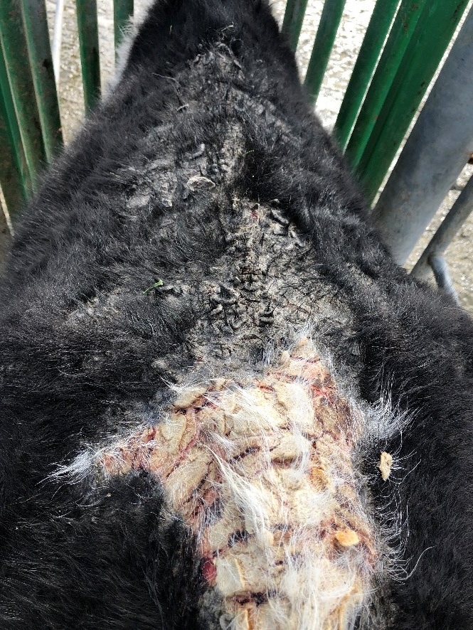
Three smears were received from skin scrapings from cattle with diffuse skin lesions. The lesions were widely distributed but were particularly severe on the white areas of the skin (Figure 1).
Laboratory findings:
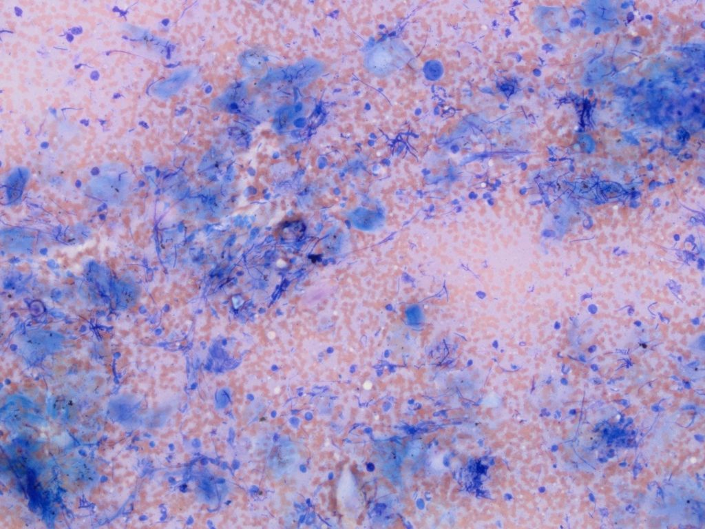
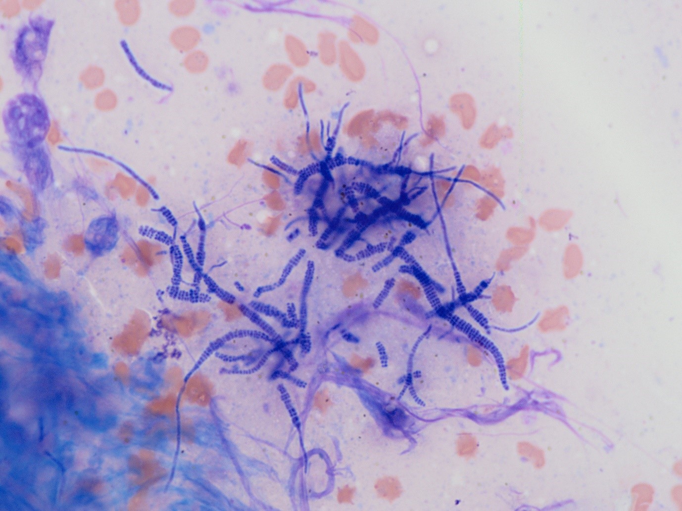
On cytology, the cellularity was high in all the smears and consisted of keratinocytes in a background of erythrocytes. Moderate numbers of inflammatory cells were seen including non-degenerate and degenerate neutrophils, lymphocytes and fibroblasts. Large numbers of bacteria with a “railway track” morphology were seen (Figure 2).
Laboratory diagnosis:
The distinctive bacterial morphology indicates infection with Dermatophilus congolensis – commonly referred to as rain scald.
Discussion:
Dermatophilosis has a worldwide distribution but is most prevalent in tropical and subtropical regions, associated with moist, humid conditions. It affects a wide range of domestic and a wild animals. In New Zealand, most cases are in cattle, sheep and cervids, but it has been described in cats and in humans.
Members of the genus Dermatophilus are aerobic, gram-positive, branching, filamentous rods. They produce motile zoospores, and aerial mycelia are ordinarily absent. Septa are formed in transverse and horizontal planes and give rise to parallel rows of coccoid cells, often referred to as “railroad tracks.” These organisms are catalase positive and non–acid fast. Dermatophilus is an obligate parasite so contact between animals is a prerequisite for infection. However, the organism can sporulate and the spores are resistant to desiccation and cause new infections when moist conditions return.
Dermatophilosis is characterised by the development of an exudative dermatitis followed by scab formation, alopecia and thickening of the skin. The serous exudate leads to the development of scabs that finally become hard, horny and confluent and they may have an almost wart-like appearance.
The two most important predisposing factors are skin damage and moisture. There was a history of facial eczema on this farm and the predominance of lesions on the white patches suggests that this may have been the initiating factor combined with the wet weather.