
AMY WEEDEN
Clinical history
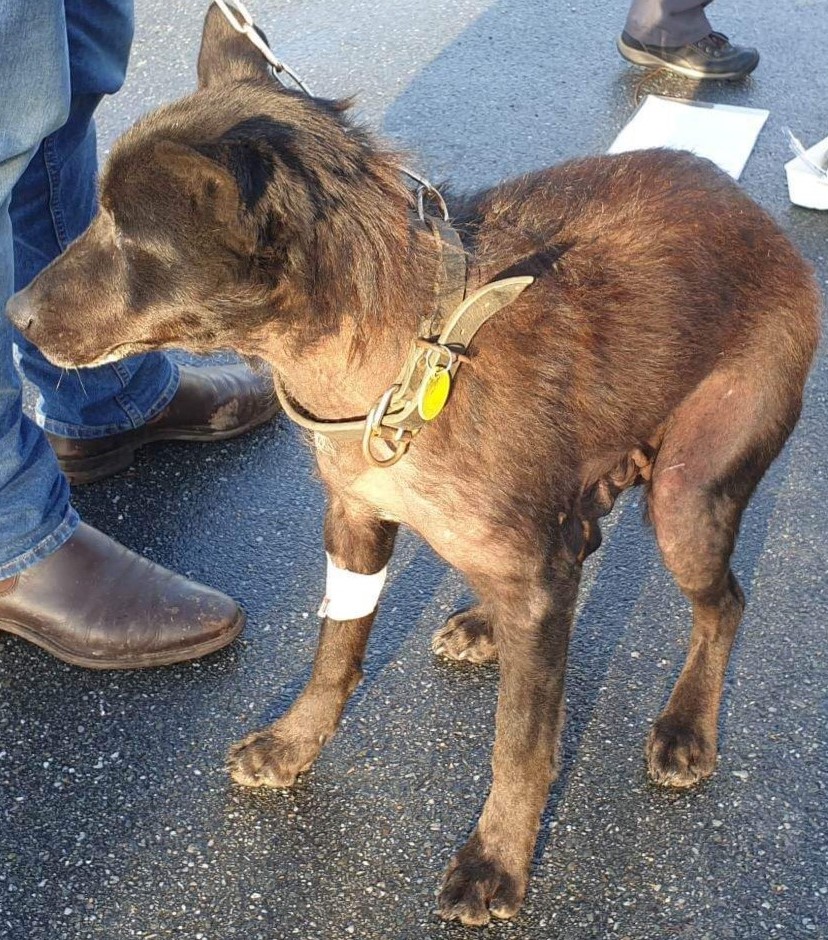
A 9-year-old male Kelpie presented with a urinary tract infection, generalised alopecia, and pendulous nipples (Figure 1). A cystic prostate and an intra-abdominal mass (possibly consistent with a testicle) were identified on ultrasound. The dog had been adopted as adult with no surgical history and had a small scrotum with no descended testicles and no obvious castration scar.
Cytology findings
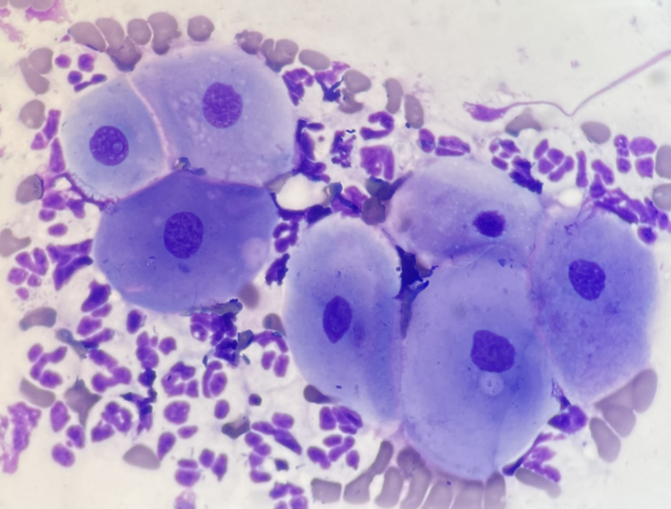
Cytology smears from the cystic prostate (Figures 2 and 3) and mass (Figure 4) were received in the laboratory.
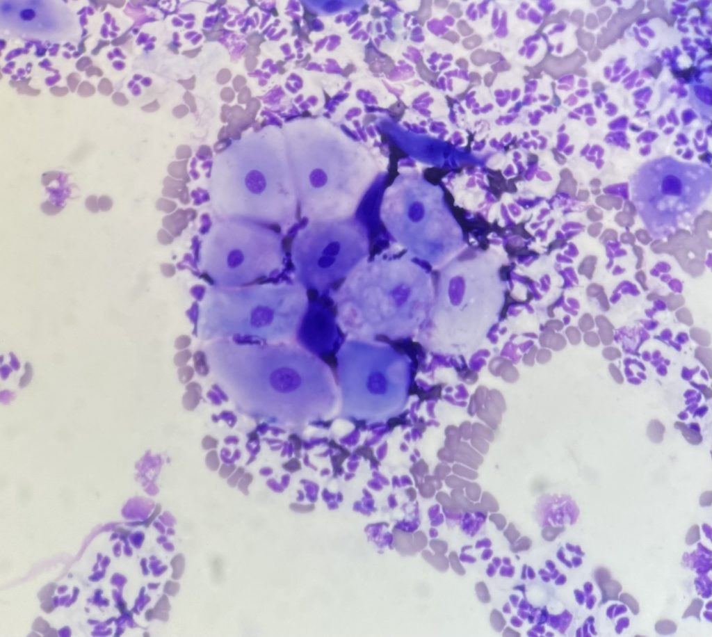
The prostate sample was mildly blood contaminated, well preserved, and highly cellular. There was a predominance of non-degenerate and variably karyolytic neutrophils, and rare macrophages were observed. There was also a population of well-differentiated to mildly atypical squamous epithelial cells, seen individually or in small aggregates. Bacteria with mixed morphology were present in moderate number, frequently found within neutrophils. Normal, cuboidal to columnar prostatic epithelial cells were not identified. Mixed growth with a predominance of Haemophilus haemoglobinophilus was isolated on culture.
The mass sample was of low intact cellularity. This sample appeared to contain a mixture of cell types, including clustered spindloid cells (probable Leydig cells), individualised discrete round cells (germ cells), and large round-to-spindloid cells with vacuoles (probable Sertoli cells) shown in figure 4) .

The prostatic cytology was consistent with squamous metaplasia and associated bacterial infection.
Discussion
Metaplasia is the transformation of one mature/differentiated cell type to another. Squamous metaplasia of the prostate is a known sequela to hyper-oestrogenism in male dogs. This is most commonly associated with Sertoli cell tumours, though administration of exogenous oestrogens could also cause this change. Chronic inflammation is another general cause of metaplasia.
Since this dog had an intra-abdominal mass and an unknown surgical/neuter history, the aspirated mass with multiple cell populations was expected to represent a testicular tumour. Based on cytology alone, a definitive diagnosis of Sertoli cell tumour could not be made, but in context of the clinical presentation, this was the working diagnosis.
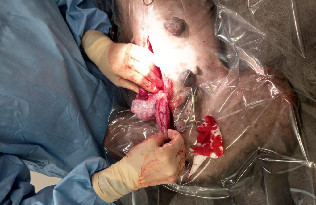
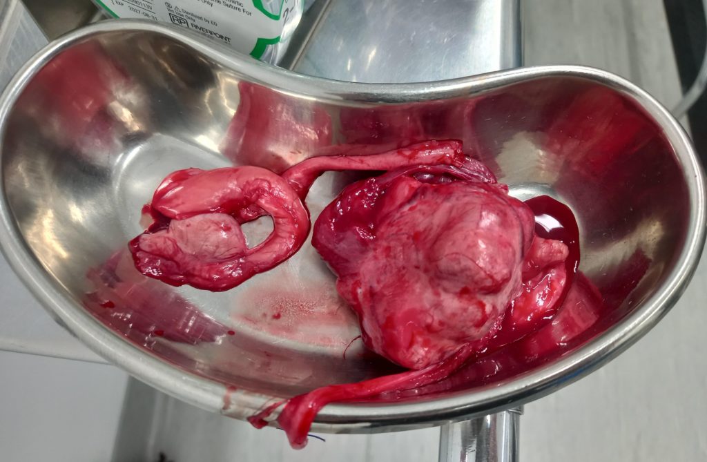
Exploratory laparotomy was performed, and two intra-abdominal testicles were identified. One of the testicles was significantly enlarged and abnormal in shape, consistent with neoplasia (Figures 5 and 6). A Sertoli cell tumour was confirmed with histopathology.
Many thanks to Rebecca Pirie at Vetlife Alexandra for submission of this excellent case and for the great images and follow-up information.
(Refer to “Very ballsy” for a related discussion on testicular tumours)