
CRISTINA GANS
It’s becoming more common in recent years for pathologists to issue a histology report which recommends the use of immunohistochemistry (IHC). I’ve frequently been asked: what is IHC and why do we need it?
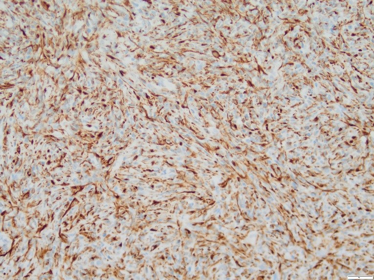
Immunohistochemistry is a molecular technique which can be used for the diagnosis and prognosis of specific tumours and for the identification of infectious agents. It employs the use of target antibodies to identify a specific antigen or “marker” in the tissue in the question, and is performed on formalin-fixed tissue. Antibodies applied to a fixed tissue are bound to a specific enzyme, which can induce a colour reaction when they bind to the specific antigen. The tissue is then viewed microscopically to determine the presence (or absence) of a colour reaction, which can indicate whether a specific antigen is present in the tissues (see Figure 1).
Although many tumours may be diagnosed through histopathology, some tumours may be poorly differentiated or show conflicting histological features which are not typical of any tumour. Immunohistochemistry may provide a definitive diagnosis in certain tumours or when a definitive diagnosis cannot be achieved, IHC may help to narrow down the list of differential diagnoses. For example, the markers cytokeratin and vimentin may be able to differentiate between a poorly differentiated carcinoma and sarcoma.
In some cases, IHC may be able to further characterise the nature of the tumour to provide information regarding prognosis. For example, B- and T-lymphocyte markers can be used to immunophenotype lymphomas, which can have prognostic and therapeutic implications.
At Gribbles Veterinary, the pathologist will be able to provide guidance to decide which markers may best for your submission. In most cases, IHC can be performed on tissues that have already been submitted, without the need for further patient sampling. Tissue blocks are sent to a referral laboratory, with a turnaround time of approximately 1 to 2 weeks.
IHC is currently only available for cats and dogs, and pricing can be found in our current price book. In some cases, some antibodies are not available in New Zealand, and we can submit samples to a referral laboratory in the U.S.A. (at an additional cost).
Reference:
Ramos-Vara, J. A. “Technical aspects of immunohistochemistry.” Veterinary Pathology. 42: 405-426, 2005.