
SUNAO FUJITA
Clinical history
A 15-year-old spayed female, domestic short haired cat presented with a large granulomatous-type lesion on the right upper lip. Fine needle aspiration of the mass and biopsy were performed, and the smears and fixed tissue were sent to the laboratory for cytology and histopathology.
Cytologic findings
Large numbers of round to sometimes spindloid cells were observed on the slides stained with Diff-Quik stain. The cells were arranged in clusters. Their nucleus was round to occasionally indented in shape and contained finely to coarsely stippled chromatin with single to multiple variably sized sometimes angular nucleoli. These cells had moderate amounts of basophilic cytoplasm with moderate N/C ratios and showed moderate anisocytosis and anisokaryosis. Sometimes bi- to multi-nucleated cells and bizarre mitotic figures were noted. The cells rarely contained scattered dark green to black intracytoplasmic pigment, morphologically most consistent with melanin (figure 1).
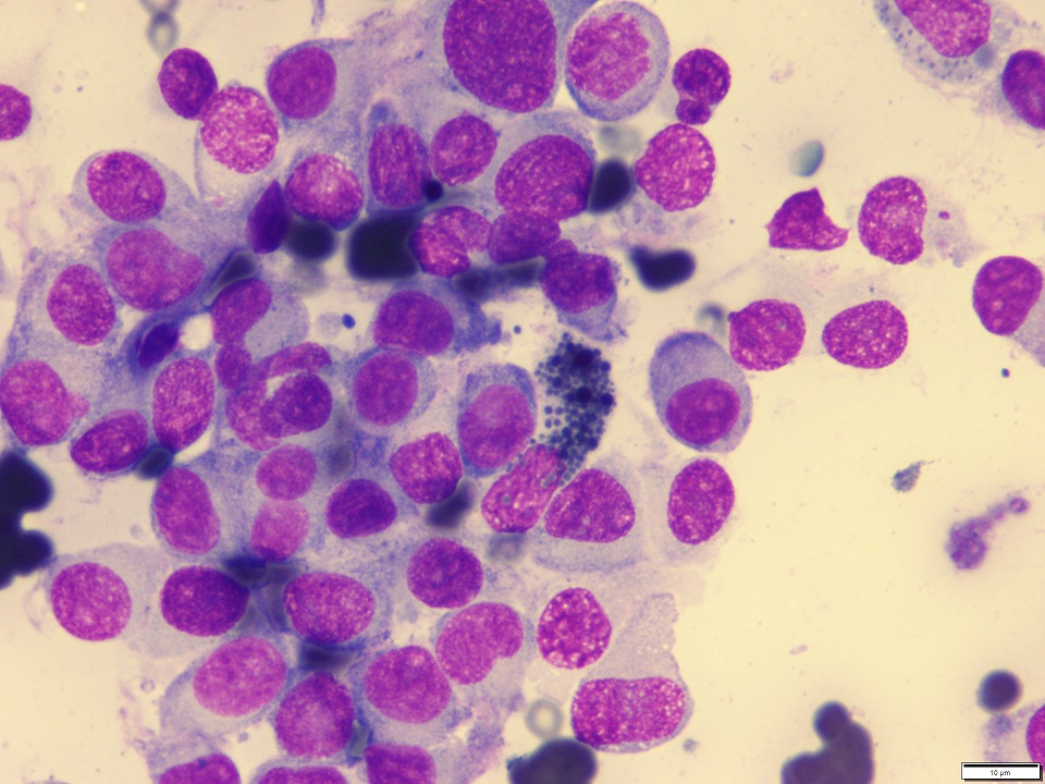
These cytologic findings suggest an anaplastic neoplasm. Given the intracytoplasmic granules, an amelanotic melanoma was suspected.
Histopathologic findings
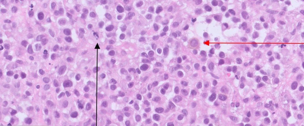
The biopsy samples were composed of fragments of a densely cellular neoplasm. The neoplasm consisted of nests and bundles of round to spindloid cells. The cells had moderately abundant pale eosinophilic cytoplasm, large round to oval nuclei with finely dispersed chromatin, and large prominent central nucleoli. There was moderate atypia characterized by nuclear hyperchromasia, karyomegaly, and anisokaryosis. The mitotic rate was high at 44 per 10hpf equivalents. Atypical mitoses were observed. Scattered cells contained small dustings of pale brown cytoplasmic pigment (figure 2).
Diagnosis
Melanoma.
Discussion
Cytologic diagnosis of melanocytic tumour is often straightforward but can be confounded when the cells lack characteristic intracytoplasmic melanin. In this case, most of the neoplastic cells do not contain
melanin pigment, which was problematic for cytologic confirmation of melanoma but the presence of rare cells with melanin was suspicious for a poorly differentiated (amelanotic) melanoma. Amelanotic cells may appear vacuolated and clear cell or balloon cell variants can have large amounts of clear cytoplasm on cytology samples, which have been reported in dogs and cats1.
Unlike dogs, melanomas are uncommon in cats, and usually uveal or cutaneous with rare oral melanomas are seen. Cutaneous melanomas account for 0.5% of all skin tumours and are commonly seen on head, tail, distal extremities, and lumbar region2. No sex predisposition is noted3. The mean age of cats with melanoma is 7.9 years (range 4.0-16.0 years)3. Cats with auricular melanomas are significantly younger (median age 7.9 years) than cats with cutaneous non-auricular melanomas (median age 13.3 years)3. It has been implicated that UV light may affect development of auricular melanomas3.
Most cutaneous melanomas are malignant in cats. Generally, the prognosis is poor due to recurrence and regional metastasis in up to half the cases2. Metastatic rates have been reported to range 5-30% in feline non-ocular melanomas3. Nonocular melanomas exhibit a wide variation in biological behaviour and predicting clinical outcomes is challenging with no widely accepted prognostic criteria. Some studies have suggested prognostic factors including low pigmentation, mitotic index (4-5 per 10 HPF), tumour size, mucosal location, and intra-tumoral necrosis3, 4, 5. Amelanotic features are most commonly considered to be associated with a poor prognosis3. One study has reported that while median survival time for cats with pigmented melanomas is 179 days, cats with amelanotic melanoma has median survival time of 71 days3.
Surgical excision is a primary option of treatment for non-ocular melanomas in cats. It has been reported that median survival time for cats treated by surgery is 143 days, but others without surgical treatment has a shorter median survival time (71 days)3.
In this case, poor pigmentation, high mitotic index, and nuclear atypia were confirmed and warranted a guarded prognosis. The cat was euthanised at approximately 3 weeks after histologic diagnosis.
Melanoma should be included in differential list when cytology reveals a neoplastic lesion composed of round to spindloid cells that do not contain melanin pigment.
Acknowledgements to Dr Michelle Ross from Manley Vet Hospital for this interesting submission.
References
1. Michael H. Melanoma. In: Veterinary cytology (Sharkey, L. C. eds.), pp. 158-165, WILEY Blackwell, Hoboken. 2020.
2. Smith SH, Goldschmidt MH, McManus PM. A comparative review of melanocytic neoplasms. Vet Pathol. 39: 651-678, 2002.
3. Chamel G, Abadie J, Albaric O, et al. Non-ocular melanomas in cats: retrospective study of 30 cases. J Fel Med Surg. 19: 351-357, 2017.
4. Pittaway R, Dobromylskyj MJ, Erles K, et al. Nonocular melanocytic neoplasia in cats: Characterization and proposal of a histologic classification scheme to more accurately predict clinical outcome. Vet Pathol. 56: 868-877, 2019.
5. Sabattini S, Renzi A, Albanese F, et al. Evaluation of Ki-67 expression in feline non-ocular melanocytic tumours. BMC Vet Res. 14:309, 2018.