
Sandy Weltan
Clinical history
A 10-year-old male, neutered, Turkish Angora cat presented with a soft but uneven textured lobulated subcutaneous mass on the rump. There was a history of a cat fight. Fine needle aspirates were submitted for evaluation.
Laboratory findings:
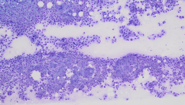
On initial examination on low power (Figure 1) it looked like a mesenchymal tumour, but what are the negatively staining bodies?
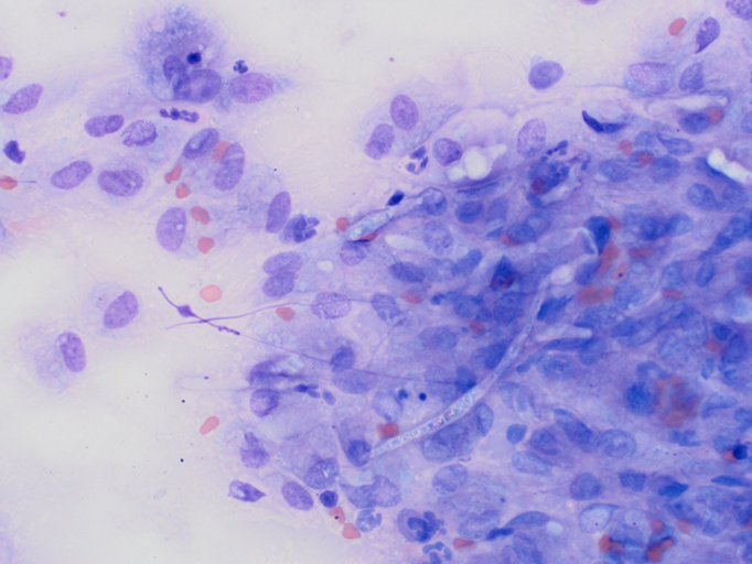
Large numbers of fungal hyphae amidst a moderately basophilic background containing large numbers of macrophages and small numbers of non-degenerate neutrophils and lymphocytes (figure 2).
The referring veterinarian was keen to perform excision of the lesion with submission for histopathology and fungal culture but unfortunately the owner declined.
Discussion:
Localised opportunistic fungal infections may result in cutaneous or subcutaneous lesions. Phaeohyphomycosis includes fungi with melanised cell walls and include genera Alternaria, Bipolaris, Exophiala, Cladophialophora, Cladosporium, Curvularia, Fonsecaea, Muyocopron, and Phialophora, among others.
They present as nodular or ulcerated lesions in areas in contact with soil, for example nose and digits. In this case there was a history of a cat fight which would have provided a portal of entry. The pigmentation is not always obvious.
Halohyphomycosis includes e a collection of ubiquitous saprophytes with hyaline cell walls, such as Acremonium, Chrysosporium, Fusarium, Geomyces, Lomentospora, Oxyporus, Paecilomyces, Penicillium, Scedosporium, Rasamsonia, and Talaromyces spp., among others. Halohyphomycosis tends to occur more commonly in dogs than in cats and often causes systemic disease.
References:
· Barrs VR et al. Invasive Fungal Infections and Oomycoses in Cats: 1. Diagnostic approach. J Feline Med Surg. 26:1098612X231219696, 2024.
· Dedeaux A et al. Opportunistic Fungal Infections in Small Animals. J Am Anim Hosp Assoc. 54:327-337, 2018.
· Dehghanpir SD. Cytomorphology of Deep Mycoses in Dogs and Cats. Vet Clin North Am Small Anim Pract. 53:155-173, 2023.