
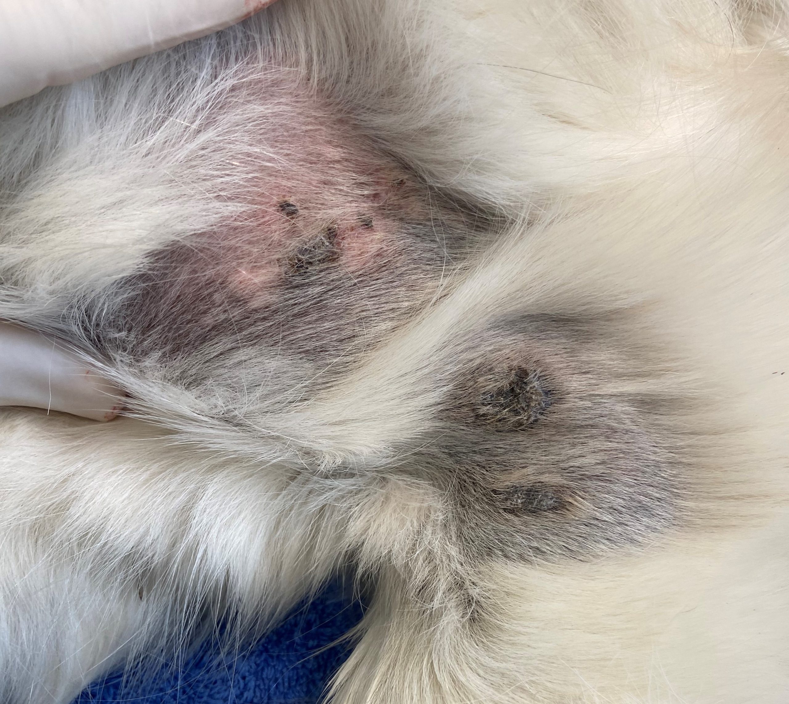
Main differential
Pemphigus foliaceous.
The cytology indicated inflammation (neutrophilic and eosinophilic) and low numbers of keratinised epithelial cells. There were also rare round cells, with deeply basophilic cytoplasm and large round to oval nuclei with a prominent nucleolus, typical of acantholytic-type cells (arrow). These findings were suggestive of autoimmune skin disease. Other differentials for the cytology findings included pustular dermatophytosis or superficial bacterial pyoderma.
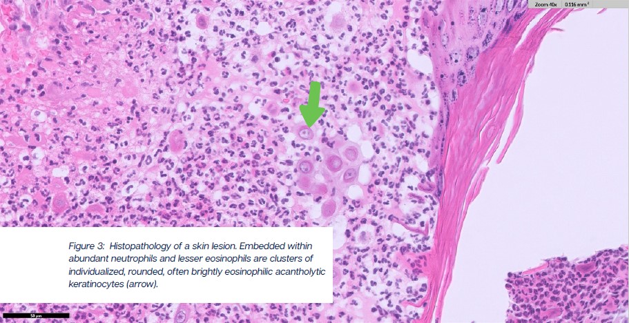
The histopathology revealed a neutrophilic and eosinophilic pustular dermatitis with acantholysis. These finding were consistent with a diagnosis of pemphigus foliaceus.
Pemphigus are autoimmune diseases where the body produces antibodies against keratinocytes desmosomal proteins, which leads to loss of cell-to-cell adhesion. Pemphigus foliaceus (PF) is the most common variant and is characterised by intra-epidermal pustules, which can be localised in the spinous, granular, or corneal layer.
The autoantibodies of PF target the extracellular component of some cadherins that make up desmosomes. After the breakage of intercellular junction, neutrophils will fill the intra-epidermal cleft and give origin to the non-follicular pustules.
These will rapidly dehydrate and develop into erosions, crusts, scales, and alopecia.
Cause and predisposition
In dogs and cats, most cases of PF are presumed to be idiopathic. However, a possible association between allergic skin diseases or exposure to drugs and development of PF has been reported. Reported drugs associated with PF include antibiotics and selected topical ectoparasitic preparations. Other potential associations include concurrent systemic disease and sunlight exposure.
Breed predisposition in dogs is observed with the idiopathic and the drug induced form of the disease.
The drug induced form is more often seen in the Doberman pinscher and Labrador retriever. A genetic predisposition for idiopathic PF is noticed in bearded collie, akita, chow chow, Newfoundland, schipperke, and Doberman. In addition, English springer spaniel, Chinese shar pei, and collie also have been shown to be at increased risk. The facially predominant form is most seen in chow chows, and akitas. No strong cat breed predisposition to PF has been reported; however, the most represented breeds are domestic shorthair, medium-hair, and longhair cats.
Although PF is typically a disease of middle-aged dogs, any age can be affected. For dogs and cats, predisposition to PF does not seem to be associated with the animal’s sex.
Clinical signs
The primary lesion in patients with PF is the pustule. However, because most PF pustules are superficial, small, and therefore transient, identification of pustules can be a rare and challenging opportunity. Most pustules will instead rapidly evolve into erosions and crusts. Indeed, crusts are the most common clinical presentation of PF in dogs and cats and will often appear thick (multilayered) with a yellowish coloration due to the cyclic nature of PF. Other lesions include pustules of variable size, erosions, and alopecia. The lesions can coalesce or cluster and organize into different patterns. Pruritus is variable but commonly reported and can be severe in patients of both species.
The most common areas involved are dorsal muzzle, nasal planum, pinnae, periorbital skin and paw pads. A striking bilaterally symmetric distribution of clinical lesions is a key feature of this condition.
A notable distinction between PF in dogs and in cats is the presence of lesions in the ungual folds and periareolar region of cats. Periareolar lesions lead to high suspicion for feline PF, although lesions are less commonly found in this area than in other areas. A relatively high number of dogs and cats also exhibit systemic signs of lethargy, pyrexia, hyporexia or anorexia, weight loss, and pain.
Diagnosis
Clinical suspicion of PF should arise from the classic clinical features of superficial pustular dermatitis. The next recommended diagnostic step for cases with suspicion of PF is cytology to detect inflammation with acantholytic keratinocytes.
The cytology often reveals numerous nondegenerate neutrophils. About half of the canine cases also have eosinophils. Acantholytic keratinocytes have reportedly been observed in cytologic samples from approximately 2/3 of dogs and cats. However, presence of acantholytic keratinocytes is not pathognomonic for PF because pyoderma, dermatophytosis caused by Trichophyton species, and canine leishmaniasis (not endemic in New Zealand) can also produce acantholytic keratinocytes. Surgical biopsy and histopathological examination of the skin remain the gold standard for diagnosis of pemphigus foliaceus. Pustules are the most informative lesions in these patients. Thoroughly check commonly affected areas. If pustules are not seen, then select thick crusts. It is very important not to surgically scrub or close-clip areas before histology biopsies, as this will remove the crusts and result in a missed diagnosis.
Conclusion
In summary, pemphigus foliaceus should be considered in dogs and cats presenting with superficial pustules, erosions, crusting, alopecia, and lesions with a bilaterally symmetrical distribution, particularly involving the face, pinnae, nasal planum, and paw pads. Cytology is a valuable first-line diagnostic tool, as the identification of neutrophilic inflammation with acantholytic keratinocytes can strongly support clinical suspicion. However, because acantholytic keratinocytes are not pathognomonic, histopathology remains essential to confirm the diagnosis and distinguish PF from important differentials such as superficial pyoderma and dermatophytosis.