
Species: Canine**, feline**, equine (**please see limitations below)
Specimen: 2 mL serum
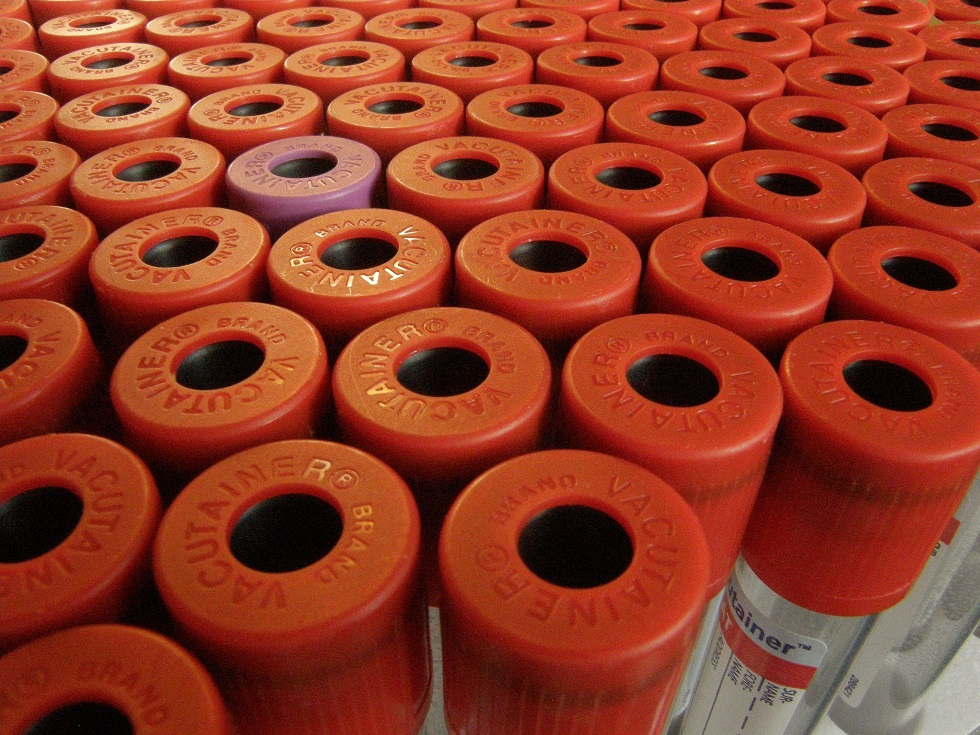
Container: Plain (red top) or gel tube
Collection protocol: Standard venepuncture
Special handling/shipping requirements: Samples should be transported to the laboratory within 24 hours
General information about the disease: N/A
General information about when this test is indicated:
Determination of gonadal status of an animals in circumstances including:
- Suspected cryptorchid males
- Female cats and dogs with unknown desexing history, particularly stray animals
- After desexing surgery to confirm complete removal of gonadal tissue**
- Supposedly desexed bitches and queens exhibiting signs of oestrus (confirmation of “ovarian remnant syndrome”)**
Anti-Müllerian hormone (AMH) is produced by the follicles of a sexually mature ovary and Sertoli cells in a sexually mature testes. After complete ovariectomy or castration, levels of AMH decrease significantly. Intact bitches and dogs, and cryptorchid animals, will have higher levels of AMH than completely desexed animals. A single serum test can differentiate these animals. After desexing, it is recommended to wait at least 30 days before testing, when evaluating for the presence of functional gonadal tissue. In general, low levels of AMH indicate the animal is desexed, high levels indicate the animal has functional gonadal tissue.
**It is important to note that data are lacking concerning animals with ovarian remnant syndrome. One study indicates that AMH is reliable for detection of ovarian remnant syndrome, but this study also points out that accuracy may be assay-dependent (Turna 2015). A positive AMH result in an animal showing signs of retained ovarian tissue helps with confirmation (ruling in). However, a negative AMH does not completely exclude/rule out the presence of retained gonadal tissue. If a negative AMH result is reported in an animal with clinical signs of retained ovarian tissue, additional diagnostics (e.g. vaginal cytology, progesterone, imaging, or a gonadotroph stimulation test) should be pursued.
Note that the assay used by Awanui Veterinary is a human assay, which has not been fully validated in veterinary species. Testing performed at Awanui Veterinary indicates that this test performs best in male horses for detection of cryptorchidism. AMH can be used to aid in the diagnosis of granulosa cell tumours in horses. However, the human assay used by Awanui Veterinary has not been evaluated for that utility, and is not recommended at this time for diagnosis of GCT.
Comparison with other related tests:
A single random blood sample for AMH testing is sufficient to distinguish a fully neutered animal from a completely intact animal. In-house methods to assess gonadal status include vaginal cytology (which requires the animal to be in oestrus) or ultrasound (which requires an experienced operator, and may miss small ovarian remnants). Serum progesterone or testosterone can be measured, but interpretation can be hampered by naturally fluctuating levels of each, particularly with stage of cycle in females and with cryptorchidism in dogs. Stimulation with gonadotrophs (hCG or GnRH) may be required to demonstrate functional gonadal tissue.
Note: This test was developed for human AMH has not been fully validated in horses and results should be interpreted along with clinical picture.
References:
- Almeida J, Ball BA, Conley AJ, Place NJ, Liu IK, Scholtz EL, Mathewson L, Stanley SD, Moeller BC. Biological and clinical significance of anti-Müllerian hormone determination in blood serum of the mare. Theriogenology. 2011, 76, 1393-403.
- Axnér E, Ström Holst B. Concentrations of anti-Müllerian hormone in the domestic cat. Relation with spay or neuter status and serum estradiol. Theriogenology. 2015, 83, 817-21
- Ball BA, Almeida J, Conley AJ. Determination of serum anti-Müllerian hormone concentrations for the diagnosis of granulosa-cell tumours in mares. Equine Vet J. 2013, 45, p199-203.
- Claes AN, Ball BA. Biological Functions and Clinical Applications of Anti-Müllerian Hormone in Stallions and Mares. Vet Clin North Am Equine Pract. 2016, 32, p451-464.
- Claes A, Ball BA, Almeida J, et al. Serum anti-Müllerian hormone concentrations in stallions: developmental changes, seasonal variation, and differences between intact stallions, cryptorchid stallions, and geldings. Theriogenology. 2013, 79, p1229-35.
- Gharagozlou F, Youssefi R, Akbarinejad V, Mohammadkhani NI, Shahpoorzadeh T. Anti-Müllerian hormone: a potential biomarker for differential diagnosis of cryptorchidism in dogs. Vet Rec. 2014, 175, p460
- Murase H, Saito S, Amaya T, Sato F, Ball BA, Nambo Y. Anti-Müllerian hormone as an indicator of hemi-castrated unilateral cryptorchid horses. J Equine Sci. 2015, 26, p15-20.
- Place NJ, Hansen BS, Cheraskin JL, Cudney SE, Flanders JA, Newmark AD, Barry B, Scarlett JM. Measurement of serum anti-Müllerian hormone concentration in female dogs and cats before and after ovariohysterectomy. J Vet Diagn Invest. 2011, 23, p524-7.
- Themmen APN, Kalra B, Visser JA, Kumar A, Savjani G, de Gier J, Jaques S. The use of anti-Müllerian hormone as diagnostic for gonadectomy status in dogs. Theriogenology. 2016, 86, p1467-1474. doi: 10.1016/j.theriogenology.2016.05.004
- Turna Yilmaz Ö, Toydemir TS, Kirsan I, Gunay Ucmak Z, Caliskan Karacam E. Anti-Müllerian hormone as a diagnostic tool for ovarian remnant syndrome in bitches. Vet Res Commun. 2015, 39, p159-62.