
JULIE TOMLINSON
Clinical history:
A 5 year old female Guinea-pig presented with weight loss and generalized peripheral lymphadenopathy. FNAs from the right inguinal and left prescapular nodes were submitted for cytology testing.
Cytological findings:
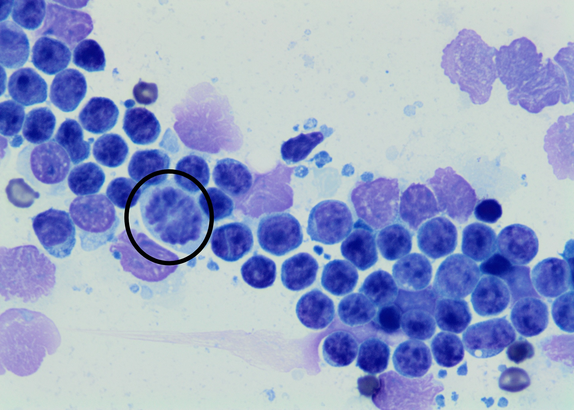
The lymph nodes consisted predominantly of intermediate sized lymphocytes admixed with few small lymphocytes, rare heterophils and eosinophils, and a small amount of blood.
Individual lymphocytes were approximately the same size or slightly larger than a heterophil. They had a round nucleus with granular chromatin, often a single round to elongate central nucleolus, and a small amount of basophilic granular cytoplasm.
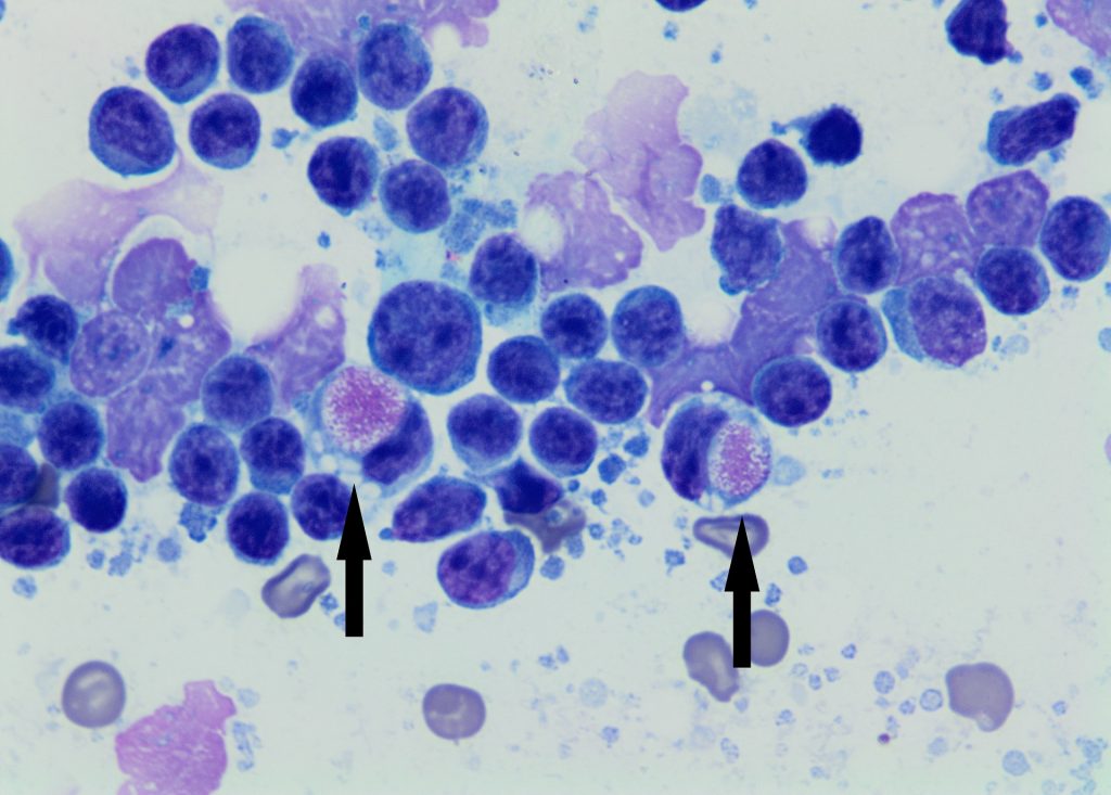
Frequent large mononuclear cells containing large variably eosinophilic cytoplasmic inclusions or Kurloff cells (see arrows) were also noted as were frequent mitotic figures (circled).
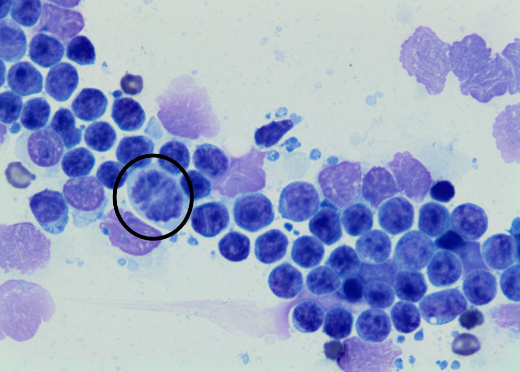
Diagnosis: Lymphoma.
Discussion:
Little information regarding lymphoma in Guinea pigs could be found in literature, but both multicentric and cutaneous lymphoma have been reported.
Kurloff cells are commonly seen in Guinea pigs and have also been reported in Capybaras. Their role is not completely understood but they are thought to have immune and natural killer activity as well as anticancer properties. Numbers are higher in females than in males and increase during pregnancy, estrus, treatment with oestrogens, and with immunologic stimulation.
Thanks to Oliver Reeve, Onewa Road Vets for this interesting case.
Reference:
Jara LF, Sánchez JM, Alvarado H, Nassar-Montoya F. Kurloff Cells in Peripheral Blood and Organs of Wild Capybaras. Journal of Wildlife Diseases 41:431–434, 2005.