

Rebecca Allan
Clinical history
An otherwise well, 9-year-old male neutered domestic medium-hair cat, presented to the veterinarian after his owner discovered he had swollen crusted toes on one front foot and both back feet. Clinical examination revealed that affected toes were swollen, malodourous, with a necrotic appearance around the nail base (photos 1 and 2). The rest of the clinical examination was unremarkable.
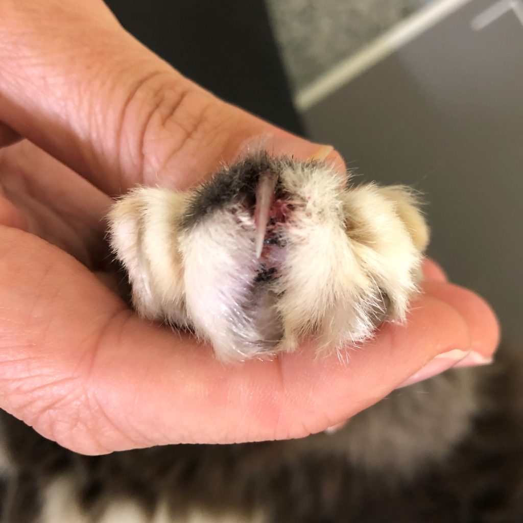
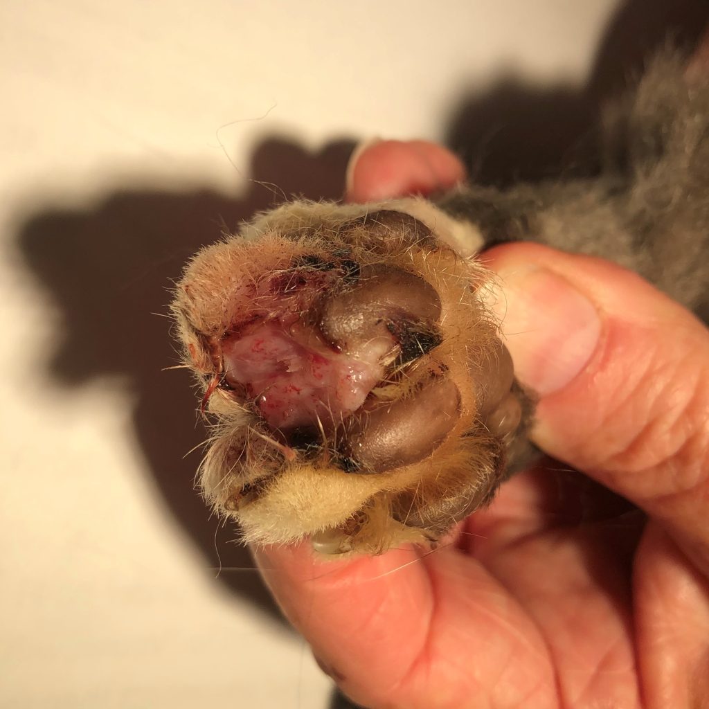
Photos 1 & 2: Affected toes are swollen and crusted.
Aspirates of affected toes were submitted for cytology, swabs of the nail beds submitted for bacterial and fungal culture and a biopsy of nail bed from one affected toe, submitted for histopathology.
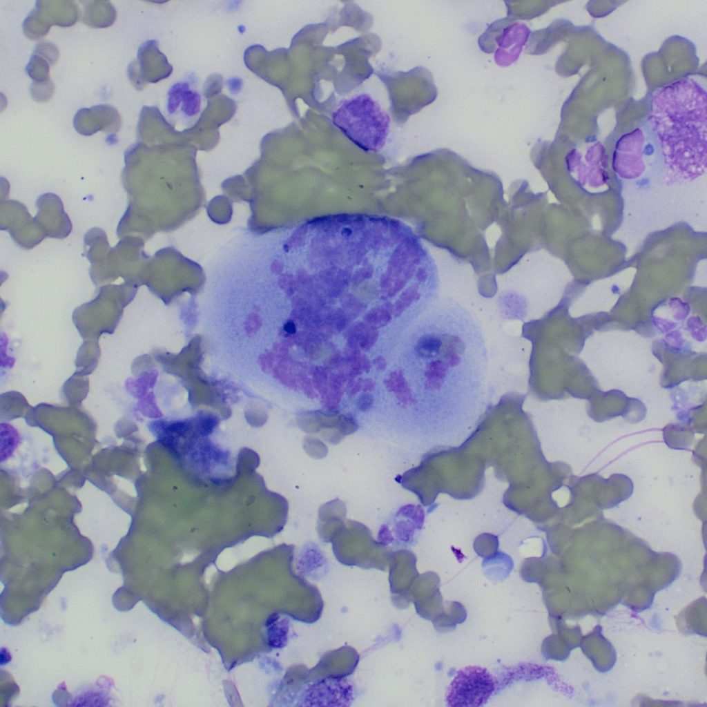
Cytology
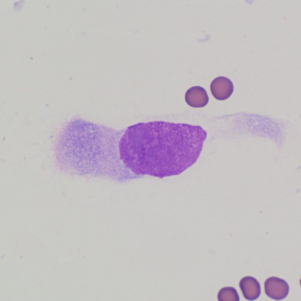
Smear examination revealed a population of epithelial cells displaying moderate to marked atypia and criteria of malignancy, including a high N:C ratio, moderate variation in cell and nuclear size (photo 3), and occasional large mitotic figures (photo 4). Rarely, a fine eosinophilic fringe was noted on the free edge of some of the cells in the denser clusters (photo 5) and occasional cells with columnar morphology were identified (photo 6).

Diagnosis
A cytological diagnosis of epithelial neoplasia was made, suspicious of feline lung-digit syndrome due to the involvement of three digits on three different limbs and presence of cells with respiratory epithelial morphology. In light of this, thoracic imaging was recommended.
Additional testing
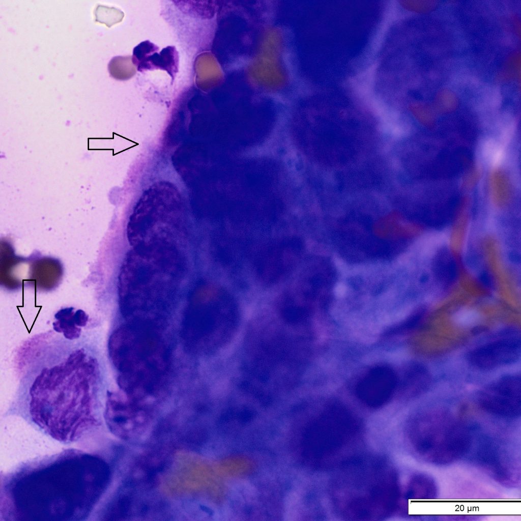
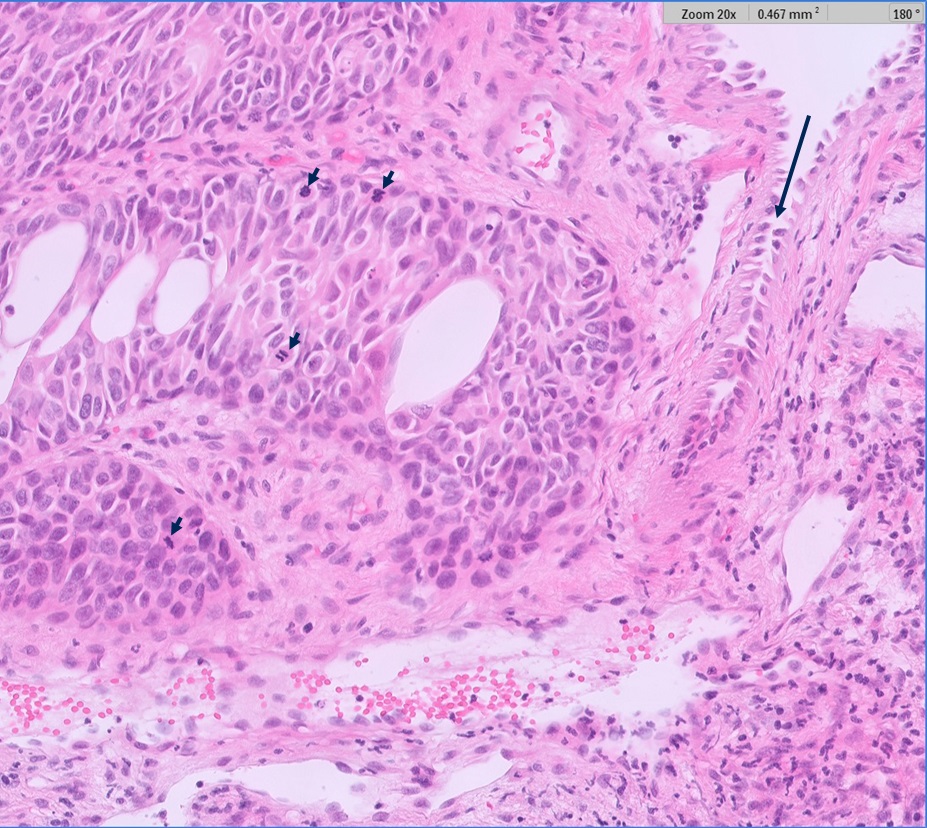
Subsequent histopathology of the nail bed biopsy revealed an infiltrative epithelial neoplasm, with marked nuclear and cellular atypia, numerous and bizarre mitotic figures and with rare cells displaying a fine ciliated border (photo 7). Results reflected cytology findings and confirmed a diagnosis of carcinoma, most likely metastatic pulmonary adenocarcinoma. Based on this news, the decision was made to euthanise the cat.
Discussion
Feline lung-digit syndrome refers to a clinical entity where primary lung tumours (often bronchogenic adenocarcinoma), which may themselves be clinically silent, as in this case, present because of metastatic lesions in one or more digits.1 Affected digits involve the most distal phalanx. Tumour emboli can also lodge in other locations causing metastatic lesions in bone, skeletal muscle, skin, eyes, and distal aorta, leading to atypical or cryptic presentations.1
Differentials to consider include bacterial paronychia (e.g. Nocardia sp. or Mycobacterium sp.), fungal infection, and immune-mediated disease.2
Primary lung tumours are not common in the cat but tend to be malignant and carry a poor prognosis. Patients with feline lung-digit syndrome, have short median survival times of only a few weeks following diagnosis. Unfortunately, digital amputation has not been shown to be palliative as further metastases rapidly develop.2
Acknowledgements
Thanks to Vetlife Richmond for the interesting case and clinical photos.
References
1. Thrift E, Greenwell C, Turner AL, Harvey AM, Maher D, Malik R. Metastatic pulmonary carcinomas in cats (‘feline lung-digit syndrome’): further variations on a theme. JFMS Open Rep. 3:2055116917691069, 2017.
2. Goldfinch N, Argyle DJ. Feline lung-digit syndrome: unusual metastatic patterns of primary lung tumours in cats. J Feline Med Surg. 14:202-208, 2012.