Saeed Sharif & David Tisdall
Parapoxviruses (PPVs; genus Parapoxvirus) infect a wide range of species generally causing localised cutaneous lesions. The genus comprises four species: Orf virus (ORFV), Bovine papular stomatitis virus (BPSV), Pseudocowpox virus (PCPV), and Parapoxvirus of red deer (PVNZ). The most common hosts of PPVs are ruminants, including sheep and goats (infected by ORFV), cattle (infected by BPSV and PCPV), and deer (PVNZ). Wildlife, including seals and sea lions can also be infected. All PPVs are known to be zoonotic, infecting humans after direct or indirect contact with infected animals.
Laboratory diagnosis
In 2022 we started developing our generic qPCR test for Parapoxviruses, it was launched as a routine test last year and this year we’ve made it even better! In response to client feedback, the molecular team has further developed the test to identify the three specific PPV species common in New Zealand:
> Orf virus (ORFV)
> Bovine papular stomatitis virus (BPSV)
> Pseudocowpox virus (PCPV)
The specific PPV qPCR is a fast, sensitive and affordable test for molecular detection and differential diagnosis of PPVs infections.
The test can be used where proliferative lesions are found on the skin or oral mucosa of suspect cases. The samples can include scab material, skin lesion or dry swabs of lesions.
Disease presentations
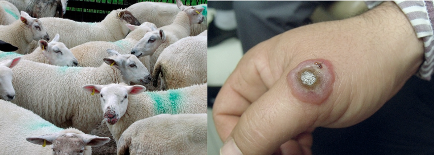
Orf is a common disease of sheep, causing papules and vesicles on the lips (‘scabby mouth’) and sometimes around the nostrils and eyes (Figure 1). Lesions can also develop on the udders when ewes suckle infected lambs. Lesions heal within about one month however infective virus can persist in dried scabs in the environment for long periods. Vaccination with a live, non-attenuated virus is practiced in some countries, including New Zealand.
Pseudocowpox occurs in dairy herds worldwide. Infection can occur on muzzles of nursing calves and on the teats of milking cows, infection occurring through small abrasions during milking or by mechanical virus transmission by flies.
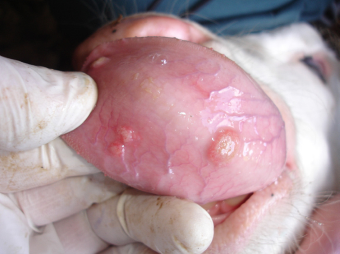
BPSV causes lesions on the muzzle, margin of the lips and buccal mucosa mainly in cattle less than two years of age (Figure 2). Suckling calves may produce lesions resembling pseudocowpox on their mother’s teats.
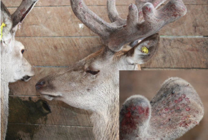
PVNZ causes scabby lesions on the muzzle, lips, face, ears, neck, and velvet, causing significant economic losses (Figure 3).