Nothing like a party on the slide!  
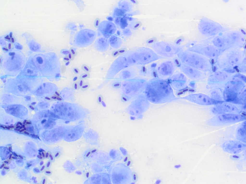
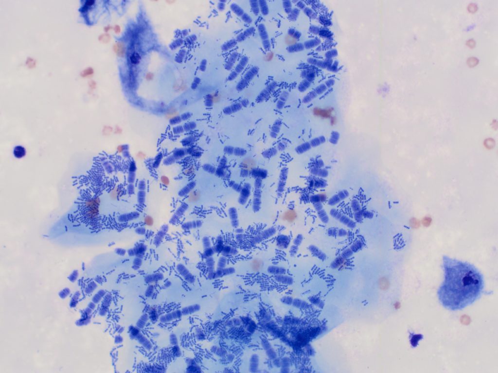
KATHRYN JENKINSIn a recent cytology smear from a cystic oral mass in a cat, we observed several clumps of large, polygonal, sky blue, keratinised squamous epithelial cells. There are a few scattered erythrocytes in the background for size comparison (see image below). An interesting finding were the mixed bacteria adherent to the squames, including numerous […]

Dogs, lymphoma and ICC

SUNAO FUJITAImmunocytochemistry (ICC) is an adjunct diagnostic tool, which helps to identify cellular origin utilizing the accuracy of antibody-antigen binding. In comparison with immunohistochemistry (IHC), which is more commonly used in both human and veterinary medicine, ICC is not routinely exploited for the classification of a variety of neoplastic lesions.However, ICC has the potential to […]

Case of the month

KATHRYN JENKINSDid you know you can submit up to six slides per site, for each cytology case?Cytology tip:Increasing the number of sites sampled from each lesion, increases the diagnostic power for a more accurate interpretation. In many cases, increasing the sample number taken, improves the chances of a diagnostic sample (often we find that the […]

Case of the month

REBECCA ALLANOften the most impressive and captivating cytology smears, are the ones that require us to deliver the worst news and this case was no exception.Clinical history:An adult male budgie presented to the veterinarian with a 15-20 mm soft tissue mass in the upper femoral area of the left leg. He was non-weight bearing and […]