An unusual cause of neurologic signs in a young cat

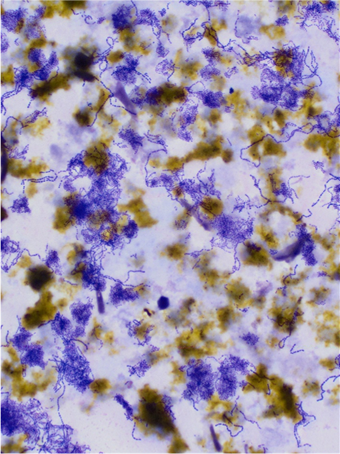
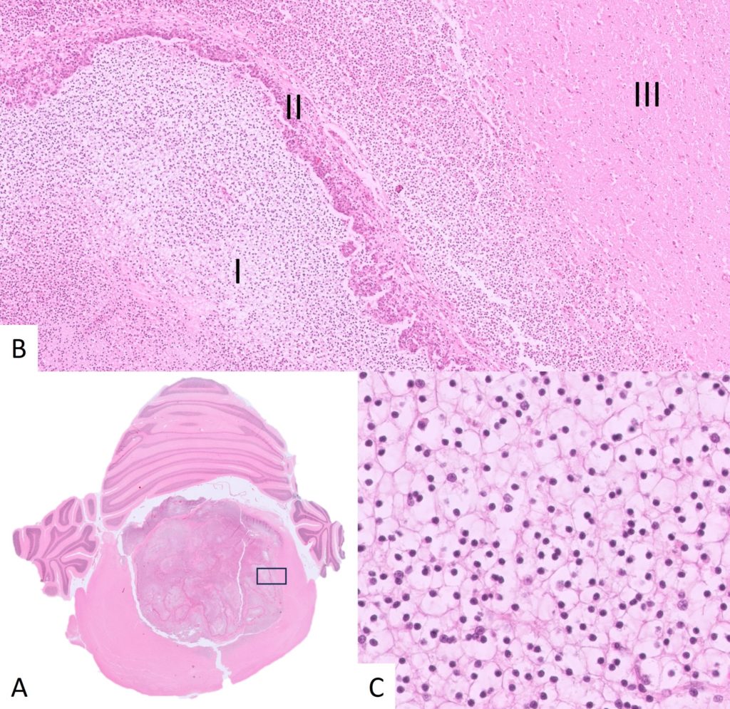
EMMA GULLIVERClinical historyA one-year-old male, neutered, domestic shorthair cat was seen by the referring veterinarian for evaluation of ataxia and a head tilt. Routine haematology and biochemistry were within normal limits, and there were no abnormalities detected on skull radiographs. The cat otherwise seemed well in himself, however clinical signs were progressive and there was […]

How galling

SANDY WELTANThe cat or dog that ain’t doing right. We all see those frustrating cases, slight lethargy, intermittent vomiting, abdominal pain, and non-specific abnormalities in biochemistry or haematology. Some may present acutely ill with jaundice and elevated liver enzymes. Tests for pancreatitis may or may not be positive and ultrasound may identify abnormalities in some […]

Lumps and bumps

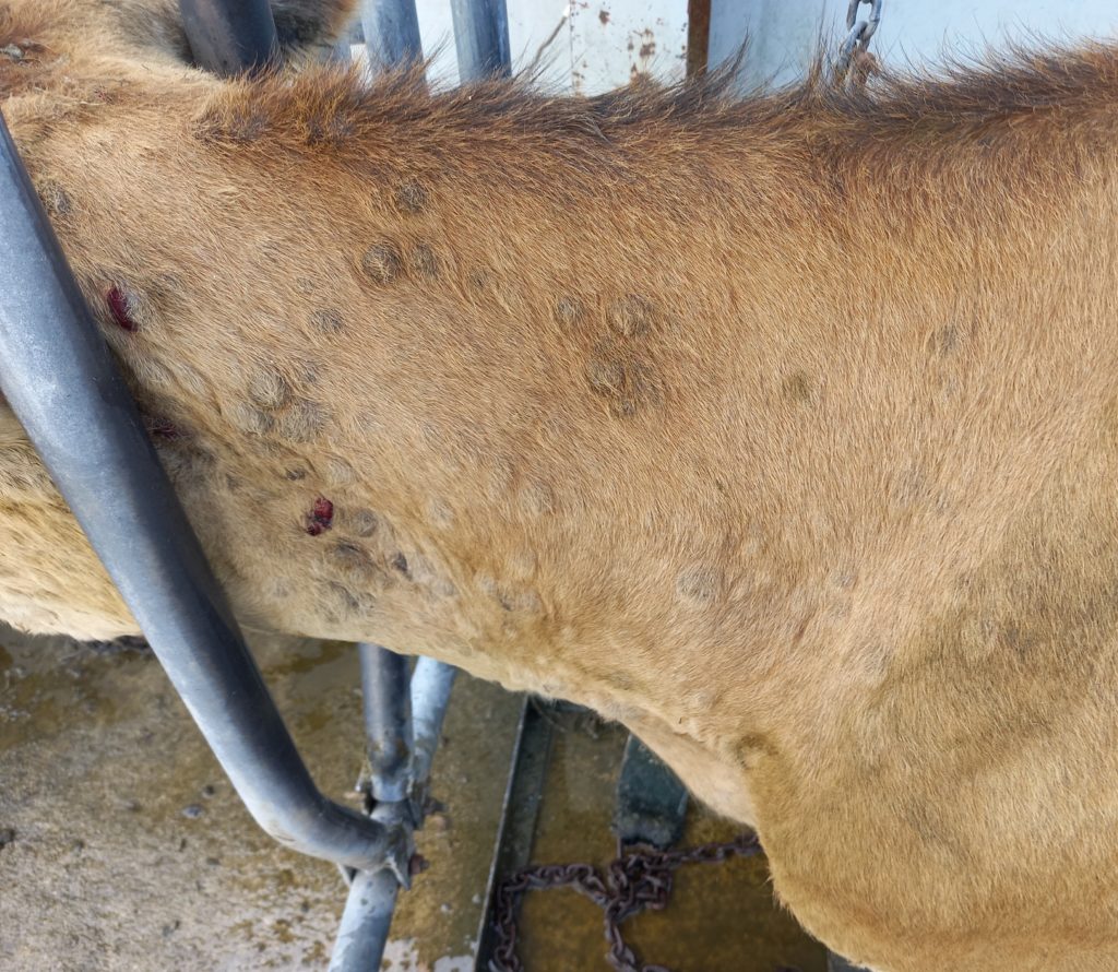
CRISTINA GANSClinical historyA two-year old Jersey cow presented to the veterinarian with multiple cutaneous nodules of unknown duration, located throughout the body, although most were concentrated on the head and neck (Figure 1). Some nodules appeared ulcerated and may have been secondarily infected. Lymph nodes were not obviously enlarged. Other animals in the herd were […]

That’s just nuts!

AMY WEEDENClinical historyA 9-year-old male Kelpie presented with a urinary tract infection, generalised alopecia, and pendulous nipples (Figure 1). A cystic prostate and an intra-abdominal mass (possibly consistent with a testicle) were identified on ultrasound. The dog had been adopted as adult with no surgical history and had a small scrotum with no descended testicles […]

Eosinophilic surprises

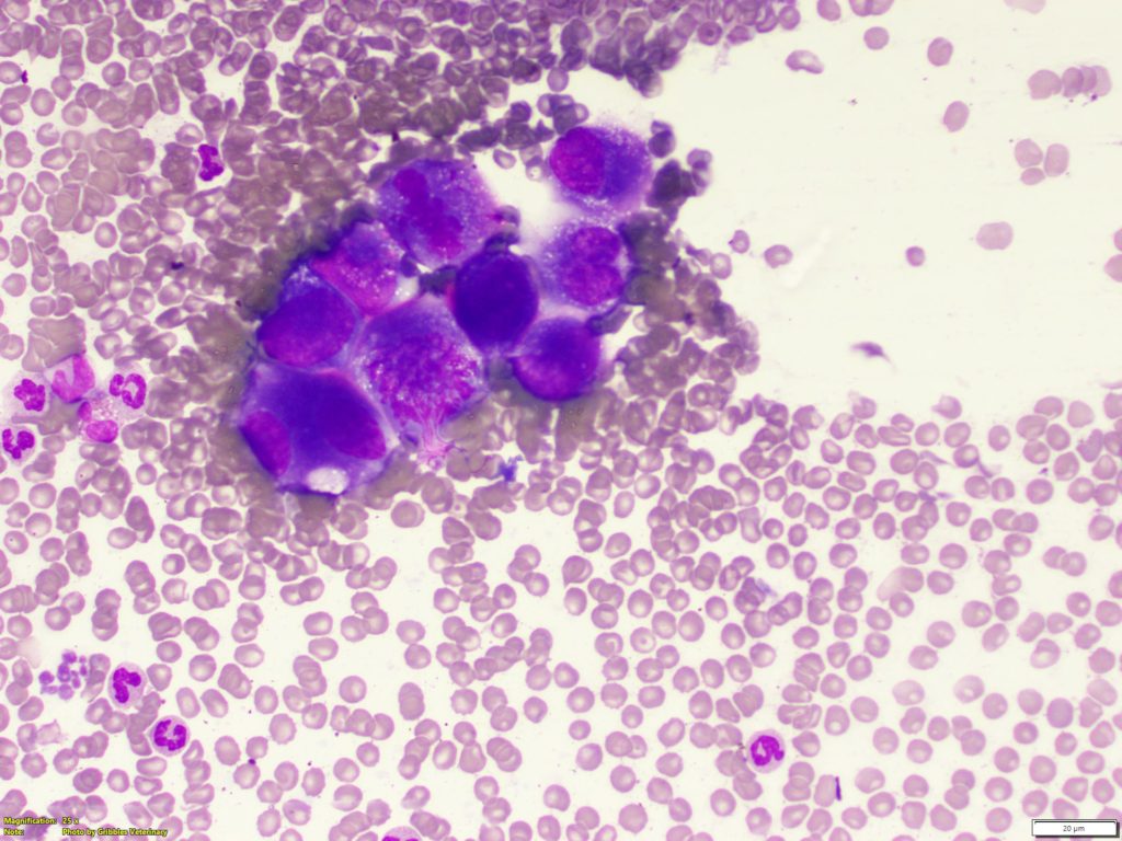
KATHRYN JENKINSOn blood smear evaluation and in cytology, eosinophils have distinctive morphological features that make them easy to identify. However, this is not always the case. Here are some surprising tips about the humble eosinophil, and a case report when all was not as it seemed on the haematology analyser results.Typical eosinophilsEosinophil granules have an […]

Salmonellosis in indoor cats

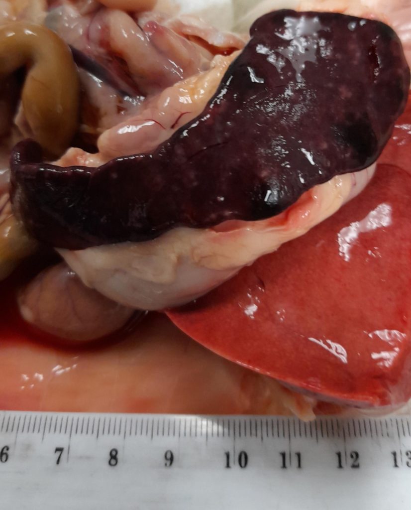
CATHY HARVEY & LISA HULME-MOIRClinical historyAfter a short period of illness (quiet and hunched, with pale mucous membranes) an approximately 6 week-old female, intact pedigree kitten died despite supportive treatment.  At autopsy the liver was enlarged with rounded margins and light brown with a prominent reticular pattern. The spleen was enlarged (90×10 to 30×5 mm) […]

Bad blood

AREFEH RAVANBAKHSHClinical historyA 12-year-old female spayed German Shepherd presented to their veterinarian for examination of a mammary mass. Approximately 11-months earlier, the patient had multiple mammary masses removed. Histological examination of the previous mammary masses had revealed mixture of benign and malignant mammary tumours.Approximately 10-months following surgery to remove the initial mammary masses, the patient […]

Bonfires and calves

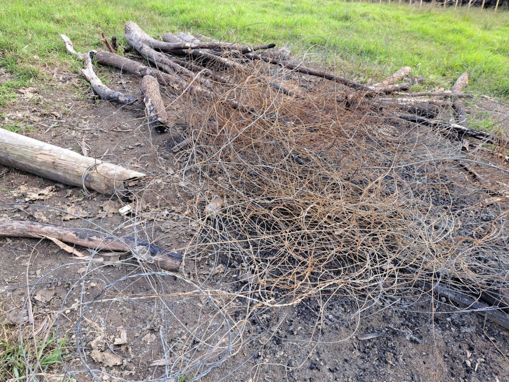
MICHAEL HARDCASTLEClinical historyThree Hereford weaners showed signs of staggering, hypermetria or recumbency and two died; one recovered with supportive care. There was possible access to cholecalciferol or cyanide, but the clinical signs were not considered consistent with those intoxications. The weaner mob had been kept in a paddock where rubbish had recently been burned.Laboratory findingsSerum […]

A little bit blue

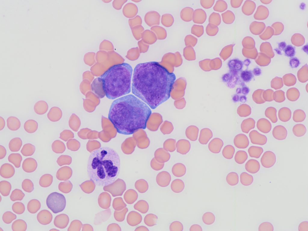
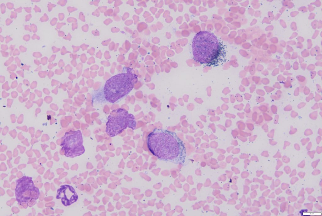
KAREN BAILEYClinical history:An adult male kororā / little blue penguin (Eudyptula minor) was brought to a wildlife hospital. On examination it was severely underweight at 805g (should be 1 – 1.2 kg) and had white mucous membranes. The PCV was 6% (normal 38-52%). An image from a blood smear (100x objective, 1000x magnification) is shown […]

It’s melanoma – and we’re not kidding!

LISA HULME-MOIRWe don’t often receive cytology samples from farm animals but just like in dogs and cats, cytology has the potential to yield a quick diagnosis that assists with decision making, optimising animal welfare and promoting judicious use of antibiotics.Clinical history:A fine needle aspirate was submitted from a lesion on the nose of a 14-year-old […]